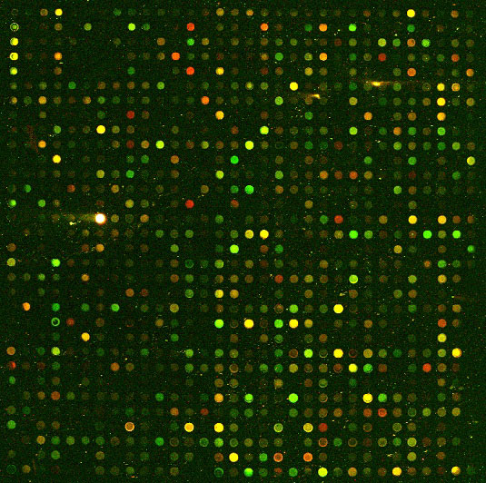
American artist Andrew Leicester has made a bit of a name for himself at Iowa State University by incorporating his art into the architectural design of the University’s Molecular Biology Building. Leicester’s much heralded G-Nome Project represents his attempt to evince the benefits and threats of genetic engineering to society, through sculpture and mosaic (1). I chanced upon Leicester’s art five years ago as an instructor at a Promega/Corning collaborative workshop in Iowa on the applicability of microarrays in transcription profiling. The workshop aimed to bring Promega’s cDNA synthesis chemistries and Corning’s cutting-edge UltraGAPS slide technologies to the attention of a small group of ebullient young scientists with a broad spectrum of research backgrounds. The floor of the atrium right outside our teaching room showcased Leicester’s ‘Novel Agents’ mosaic, incorporating a super-genetic monster as a warning of the potential perils of genetic manipulation.

Fortunately for us, the ever-prolific genomics era seems not to have spawned the terrors that Leicester depicted in his art (at least nothing as singularly destructive as a super-genetic monster). To be sure, genetic research is playing its role in the development of novel pharmaceuticals for today’s most challenging diseases. One can be similarly upbeat about the world of transcription profiling. Microarrays have come a long way since the days of self printing on a slide for single time point analysis. While valuable in their own right, microarray technologies such as those that we rolled out in Iowa give only “discrete measurements of transcriptional response” in the cells under study (2). But a robust technique called Reverse Transfection Arraying (RTA), that allows high throughput parallel transfection of tissue culture cells on a single slide, has become an attractive method for continuous monitoring of gene expression (3). Compared to conventional transfections, the order of steps is reversed- the DNA to be transfected is overlaid with cells rather than the other way around (3). But the same surface technology as that commonly employed in self printed slides (eg., GAPS, UltraGAPS) can still be used (3). Moreover microarray laboratories will already have the appropriate instrumentation and analysis software for RTA-content printing and fluorescence scanning (3,4). No further fixing or processing of transfected cells is necessary (3).
RTA has found application in siRNA inhibition studies using siRNA transfection cocktails that are spotted onto slides prior to cell overlay (4). Yet RTA is set to become a ‘cart-horse’ technique with almost limitless potential. “Studies focusing on expression changes during the cell-cycle, cell differentiation, circadian rhythms and apoptosis would greatly benefit from using living microarrays.” noted Montreal University’s Savannan Rajan in an interview with the online science magazine BioArray News (5). High-content-screening microscopy can be used to build a detailed picture of transcriptional change. Unlike 384-well plates that exhibit well-to-well variability, parallel seeding in a single chamber “increases the screening data quality” (3). Moreover the lower cell number requirement compared to traditional plating methods has made RTA the most cost-effective functional genomic technique available on the market (4). Arrays that have been printed with the desired transfection cocktail can be replicated and stored for months on end without compromising data quality (3).
Rajan has taken this technology base one monumental step further by designing a system that allows real time measurement of reporter gene expression in single cells (2). Reporters-based screening in RTA is not new. As early as 2003, scientists from UC Berkeley and Corning had worked out a way to exploit GFP to look at cell signaling, using MAPK activation as their model (6). However the single-cell applicability of Rajan’s ‘living microarrays’ make them better suited for experiments involving extreme inter-cellular heterogeneity in gene expression (2,5). In many instances researchers want to be able to see single cell differences, not just generalized patterns from a pooled population of cells (2,4). To test his system, Rajan cultured a monolayer of 293T cells over fluorescence reporter constructs that had been spotted onto chambered coverglass slides (2). This allowed uptake of the constructs into the cells and expression of the reporter, which could be detected on either a conventional microarray scanner or a fluorescence microscope (for single-cell visualization). Data was captured every 20 minutes over an experimental period of 7 days and processed with an automated image analysis system tailored for single cell segmentation (2).
Rajan’s team had to overcome some significant hurdles to ensure real time fluorescence capture (2). First, they had to identify ‘invariant features’ on the edge of each slide that could serve as positional markers since spot boundaries disappeared the moment cells were overlaid. Next, they had to get around the need to autofocus their microscopes as this would invariably expose the fluorescent reporter to damaging doses of light (2). But they did their homework and came up with a dual reporter construct called Venus-NLS (Nuclear Localization Signal)-PEST that would give them optimal brightness and most rapid polypeptide maturation. The relative amounts of material used were miniscule: 600-1000 spots of a 6.7 nl transfection complex on an 8.6 cm2 chamber slide (spots were 100–300 μm in diameter, separated by a distance of 400-1000 μm (4).
The PEST protein destablizing domain was similar to that used in some of Promega’s pGL4 vectors (2). An internal control reporter was subcloned into the same vector to avoid the stochastic variability associated with single cell co-transfections (2). Seventy five replicate spots of three drug-inducible promoters carrying a tri-copy glucocorticoid response element (GRE), cloned downstream of the adenovirus major-late minimal promoter (ADMLP) were used to test the system. And the results that Rajan’s team amassed showed unequivocally that a single-cell distribution of responses was not only achievable but could be tracked by linking photo frames over extended periods of time (2).
As my family and I drove back home from the workshop along roads that hugged the vast corn fields of central Iowa, I could not help but reflect on how the flat landscape provided a useful metaphor for thinking about advances in microarray technology. In a real sense traditional endpoint analysis gives us a similarly flat picture of gene expression that, while still informative, lacks the data richness of continuous single-cell analysis. Cells are dynamic entities that display complex responses to even the slightest external perturbations. Continuous measurements in a living microarray format are therefore crucial for accurate monitoring. In every respect, the living microarray represents the coming of age of transcription profiling.
Literature Cited
- Andrew Leicester (1991) The G-Nome Project, Molecular Biology Building, Iowa State University, Ames, Iowa
- Rajan, S., Djambazian, H., Dang, H., Sladek, R., & Hudson, T. (2011). The living microarray: a high-throughput platform for measuring transcription dynamics in single cells BMC Genomics, 12 (1) DOI: 10.1186/1471-2164-12-115
- Holger Erfle, Beate Neumann, Urban Liebel, Phill Rogers, Michael Held, Thomas Walter, Jan Ellenberg, Rainer Pepperkok (2007) Reverse transfection on cell arrays for high content screening microscopy, Nature Protocols, Vol 2, pp. 392-399
- Dominique Vanhecke and Michal Janitz (2004) High-throughput Gene Silencing Using Cell Arrays, Oncogene, Vol 23, pp. 8353-8358
- Justin Petrone (2011) Canadian Researchers Develop ‘Living’ Microarray Platform to Study Cells In Vivo, GenomeWeb BioArray News, February 22nd 2011
- Brian L. Webb, Begoña Díaz, G. Steven Martin, Fang Lai (2003) A Reporter System for Reverse Transfection Cell Arrays, Journal of Biomolecular Screening 8(6); 2003, pp. 620-623
Robert Deyes
Latest posts by Robert Deyes (see all)
- Royal Lessons for Scientific Discovery - October 28, 2016
- Because Timing Is Everything…. - August 18, 2014
- Grays On the Move: Whale Watching in San Diego - April 30, 2014
