Glowing Testimonies: A Review of NanoLuc® Use in Model Organisms

NanoLuc®

Model organisms are essential tools in the pursuit of understanding biological processes, elucidating the mechanisms of diseases, and developing potential treatments and therapies. Use of these organisms in scientific research has paved the way for groundbreaking discoveries across various fields of biology. In particular, non-mammalian models can be valuable due to characteristics such as rapid life cycles, low cost, and amenability to use with advanced genetic tools, including bioluminescent reporters such as NanoLuc® Luciferase.

NanoLuc® is a small (19.1 kDa) luciferase enzyme originating from deep sea shrimp that is 100x brighter than firefly or Renilla luciferase. It utilizes a furimazine substrate to produce its bright glow-type luminescence. In the decade following its development, the NanoLuc® toolbox has expanded to include NanoBiT® complementation, NanoBRET™ energy transfer methods, and new reagents such as the Nano-Glo® Fluorofurimazine In Vivo Substrate (FFz) which was designed for in vivo detection of NanoLuc® Luciferase, NanoLuc® fusion proteins or reconstituted NanoBiT® Luciferase. In addition to the aqueous-soluble reagents increased substrate bioavailability in vivo, with fluorofurimazine, NanoLuc® and firefly luciferase can be used together in dual-luciferase molecular imaging studies.

Here we spotlight some recent research that demonstrates how the expanded NanoLuc® toolbox can be adapted to use in non-mammalian models, shedding new light on fundamental biological processes and advancing our understanding of complex mechanisms in these diverse organisms.

Continue reading “Glowing Testimonies: A Review of NanoLuc® Use in Model Organisms”

Striking Fear into the Heart of Cardiovascular Disease Using Zebrafish and NanoLuc® Luciferase

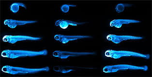
Representative images of ApoB-LP localization in zebrafish across developmental, genetic, pharmacological and dietary manipulations.
Credit: Figure 5.D of The LipoGlo reporter system for sensitive and specific monitoring of atherogenic lipoproteins by James Thierer, Stephen C. Ekker and Steven A. Farber.
Article licensed under Creative Commons Attribution 4.0 International License.

Cardiovascular diseases, or CVDs, are collectively the most notorious gang of cold-blooded killers threatening human lives today. These unforgiving villains, including the likes of coronary heart disease, cerebrovascular disease and pulmonary embolisms, are jointly responsible for more deaths per year than any other source, securing their seat as the number one cause of human mortality on a global scale.

One of the trademarks of most CVDs is the thickening and stiffening of the arteries, a condition known as atherosclerosis. Atherosclerosis is characterized by the accumulation of cholesterol, fats and other substances, which together form plaques in and on the artery walls. These plaques clog or narrow your arteries until they completely block the flow of blood, and can no longer supply sufficient blood to your tissues and organs. Or the plaques can burst, setting off a disastrous chain reaction that begins with a blood clot, and often ends with a heart attack or stroke.

Given the global prevalence and magnitude of this problem, there is a significant and urgent demand for better ways to treat CVDs. In a recent study published in Nature Communications, researchers at the Carnegie Institution for Science, Johns Hopkins University and Mayo Clinic are taking the fight to CVDs through the study of low-density lipoproteins (LDLs), the particles responsible for shuttling bad cholesterol throughout the bloodstream.

Continue reading “Striking Fear into the Heart of Cardiovascular Disease Using Zebrafish and NanoLuc® Luciferase”

Cell-free expression application: Screening for successful oligo-mediated knockdown design

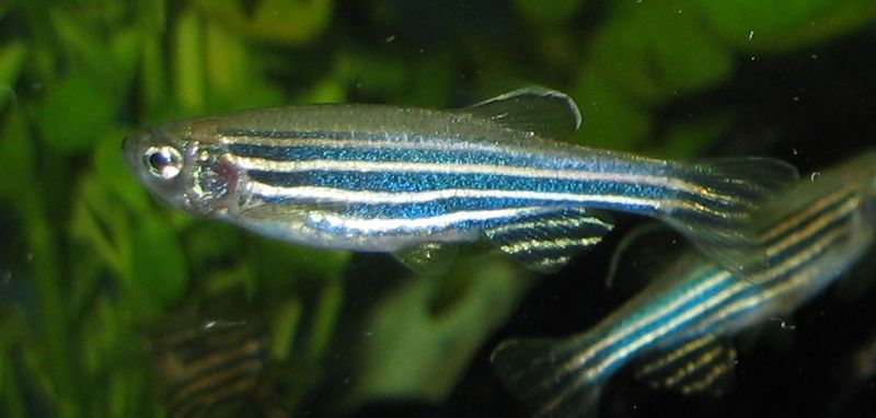
800px-ZebrafischAlthough previous references have provided data regarding the potential oncogenic role of the gene ETV7, there has been minimal investigation as to its physiological role.
In the following reference, Quintana, A. et al. (2014) Disease Models & Mechanisms 7, 265–70, zebrafish were used as in vivo model system to characterize ETV7.

One key experiment required the morpholino-oligonucleotide -mediated knockdown of in vivo ETV7. Two independent morpholinos were designed: one that inhibited translation and the other that inhibited proper splicing of exon 3. The efficacy of the translation –blocking morpholino was assessed with cell free expression of ETV7-tagged with hemagglutinin (HA).

Western blot performed with anti-HA antibodies determined the extent of the knockdown compared to a control containing no morpholino added. Once an efficient design was determined via cell-free expression screening, it was used for in vivo experiments. In conjunction additional other techniques, concluded that ETV7 is essential for normal red blood cell development.