About the Wild Life in Our Homes (at least the single-celled kind)

The initial paper from the Wild Life in Our Homes study by Dunn et al. found a correlation between the presence of dogs and specific bacterial communities on pillowcases and TV screens.

Back in the fall, I received a sampling kit, an Informed Consent form and instructions for collecting samples for the Wild Life In Our Homes citizen science project. I carefully swabbed the requested surfaces: exterior and interior door trim, kitchen counter tops, pillowcases, etc., and sent my samples in. I later received confirmation that my samples had been received and again later confirmation that they were being analyzed.

The first paper from this project has been published by Dunn et al. in PLOS ONE (Home Life: Factors Structuring the Bacterial Diversity Found within and between Homes). This initial report covers the first 40 homes sampled, all from the Raleigh-Durham, NC, USA area. Volunteers sampled their homes in the Fall of 2011, collecting specimens from nine areas: cutting boards, kitchen counters, refrigerator, toilet seat, pillowcase, door handle, TV screen, and interior and exterior door trim. The scientists used direct PCR and high-throughput sequencing to sequence the bacterial 16S rRNA gene from the submitted samples. By doing this they were able to estimate the diversity within each sample—they did not distinguish between live and dead organisms, and they did not sequence anything other than the bacterial 16SrRNA, so this study is limited to bacteria. Continue reading “About the Wild Life in Our Homes (at least the single-celled kind)”

Cold-War Bunkers Enlisted in the Fight Against Cold-Loving Fungus: More on the White-Nose Syndrome Story

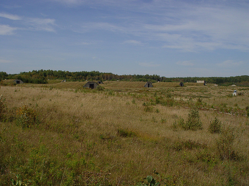
Bunkers at Aroostook National Wildlife Refuge. photo credit: USFWS/Steve Agius

A lot has happened since I first wrote about White-Nose Syndrome, the fungal disease that has devastated bat populations in North America. The disease, caused by the cold-loving fungus Geomyces destructans (now renamed Psuedogymnoascus destructans), has been identified in many more places, including most recently confirmed cases in Georgia, South Carolina, Illinois and Missouri in the United States and Prince Edward Island, Canada.

Controlling the spread of this disease is a tremendous problem, because as I indicated in a previous blog post, keeping a hardy fungus from spreading among a population of densely packed small animals in tiny, cold damp areas is not a simple task.

This problem is going to require creative solutions, and scientists at the U.S. Fish and Wildlife Service may have come up with a great idea that answers two questions: How do you control the spread of White-Nose Syndrome and what do you do with 43 unused Air Force bunkers? Continue reading “Cold-War Bunkers Enlisted in the Fight Against Cold-Loving Fungus: More on the White-Nose Syndrome Story”

The Price for Convenience May Not Be That Pricey After All

Hour glass

I was having a discussion with my mother just the other day about cleaning products (lively topic, I know). She showed me her newest time saver…prediluted bleach. Huh, I thought. I guess that does save a bit of time, but I couldn’t resist telling her that she was paying triple the price for a whole lot of water. She said, without pause, that it was worth it to her to not have to splash fully concentrated bleach around. A convenience worth paying for, in her words.

I don’t know why this struck me as odd. I pay for convenience all the time as I get older. When I started running gels back in college, I wouldn’t have dreamed of buying a precast gel, but several years into my lab life I found myself running more than 15 gels a week, so precast was really a convenient alternative. When I was a grad student, I poured all of my own plates (and most of the plates for older students, too!). Fast forward a few years, and I running upwards of 300 microbial selective cultures per week. The switch to prepoured plates was a no brainer.

When put in the context of what our time is worth, would you rather be thawing and mixing loading dyes, buffers, stains, reagents, etc., or are you better of grabbing a premixed, room-temp stable dye or ladder/loading dye mix off the shelf and getting on with your research? I think most scientists would agree that these small conveniences allow you to free up a little more time to do the important work you should be doing.

I’m curious…what time savers or convenience items do you find that make your day a little easier in the lab?

Screening for Inhibitors of CD73 (5´-ectonucleotidase) Using a Metabolite Assay

CD73

CD73 also known as 5´-Ectonucleotidase (NT5E) is a membrane-anchored protein that acts at the outer surface of the cell to convert AMP to adenosine and free phosphate. CD73 activity is associated with immunosuppression and prometastatic effects, including angiogenesis. CD73 is highly expressed on the surfaces of many types of cancer cells and other immunosuppressive cells (1). A recent study by Quezada and colleagues showed that the high concentration of adenosine produced by the CD73-catalyzed reaction on glioblastoma multiforme cells, which are characterized by extreme chemoresistance, triggered adenosine signaling and in turn, the multi-drug resistance (MDR) phenotype of these cells (2).

Because of the roles of adenosine in immunosuppression, angiogenesis and MDR phenotypes, CD73 (NT5E) is an attractive therapeutic target. However, the current methods of assaying for the ectonucleotidase activity, HPLC and a malachite green assay, are cumbersome and not suited to high-throughput screening. The HPLC assay is expensive and difficult to automate and miniaturize (3). The malachite green assay is sensitive to phosphate found in media, buffers and other solutions used in the compound-screening environment.

To address the problem of developing a reliable high-throughput screening assay for CD73, Sachsenmeier and colleagues (3) looked to a luminescent ATP-detection reagent.

Continue reading “Screening for Inhibitors of CD73 (5´-ectonucleotidase) Using a Metabolite Assay”

Proteinase K: An Enzyme for Everyone

protein expression purification and analysis

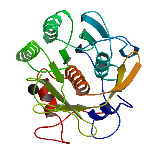
We recently posted a blog about Proteinase K, a serine protease that exhibits broad cleavage activity produced by the fungus Tritirachium album Limber. It cleaves peptide bonds adjacent to the carboxylic group of aliphatic and aromatic amino acids and is useful for general digestion of protein in biological samples. In that previous blog we focused on its use to remove RNase and DNase activities. However, the stability of Proteinase K in urea and SDS and its ability to digest native proteins make it useful for a variety of applications. Here we provide a brief list of peer-reviewed citations that demonstrate the use of proteinase K in DNA and RNA purification, protein digestion in FFPE tissue samples, chromatin precipitation assays, and proteinase K protection assays:

Continue reading “Proteinase K: An Enzyme for Everyone”

ProK: An Old ‘Pro’ That is Still In The Game

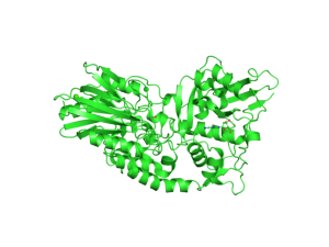
Proteinase K Ribbon Structure ImageSource=RCSB PDB; StructureID=4b5l; DOI=http://dx.doi.org/10.2210/pdb4b5l/pdb;

If you enter any molecular lab asking to borrow some Proteinase K, lab members are likely to answer: “I know we have it. Let me see where it is”. Sometimes the enzyme will be found to have expired. The lab may also have struggled with power outages or freezer malfunctions in the past. But the lab still decides to keep the enzyme. One may rightly ask – why do labs hang on to Proteinase K even when it has been stored under sub-standard conditions?

Continue reading “ProK: An Old ‘Pro’ That is Still In The Game”

Science in the Service of Art

Three artists who use science as their starting point.

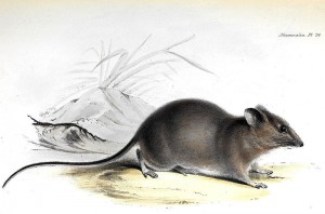
Galapagos Rice-Rat from 'The Zoology of the Voyage of H.M.S. Beagle'. This media file is in the public domain in the United States. This applies to U.S. works where the copyright has expired, often because its first publication occurred prior to January 1, 1923.
Galapagos Rice-Rat from ‘The Zoology of the Voyage of H.M.S. Beagle’ This media file is in the public domain in the United States. This applies to U.S. works where the copyright has expired, often because its first publication occurred prior to January 1, 1923.

My recent blog conversation (blogversation?) with Michele about the book The Where, The Why and The How stirred me up to think some more about the topic of science-flavored art. That book was full of delightful examples of artists using science as their inspiration; however no matter the topic or style of art, the illustrations never strayed from _illustrating_ the science they referenced. Some were more fanciful than others, but none questioned their basic intent.

Now, in literature there’s an entire genre dedicated to “science flavored” writing that ultimately doesn’t serve to illustrate any actual science concepts. I’m speaking of Science Fiction, of course, and while some early entries in the SF canon erred on the side of scientific accuracy, later practitioners of the genre took great liberties with the science, always ensuring that it served their literary goal and not vice versa. I was raised on a steady diet of Stanisław Lem books, and probably as a result tend not to demand much realism from my fiction.

On thinking about it, I’m rather surprised that the same is in general not true of art.

Continue reading “Science in the Service of Art”

“Fingerprinting” Your Cell Lines

Working in the laboratoryResearchers working with immortalized cell lines would readily agree when I state that it is almost impossible to look at cells under the microscope and identify them by name. There are phenotypic traits, however they do change with change in media composition, passage number and in response to growth factors. I remember the pretty arborizations my neuroblastoma cell line SH-SY5Y exhibited in response to nerve growth factor treatment. Thus physical appearance is not a distinguishing feature. Currently, in many labs, researchers typically use more than one cell line, and more than likely, share the same lab space to passage cells and the same incubator to grow the cells. In such scenarios, it is not difficult to imagine that cell lines might get mislabeled or cross-contaminated. For example HeLa cells, one of the fastest growing cell lines have been shown to invade and overtake other cell lines.

Misidentification of cell lines has deep and severe implications. A review of cell lines used to study esophageal adenocarcinoma found that a large number of the cell lines were actually derived from lung or gastric cancers. Unfortunately, by the time this error was discovered, data from these cell line studies were already being used for clinical trials and other advanced studies and publications. Moreover, the cell lines were being to screen and design and test specific cancer drugs which ended up in flawed clinical trials. Continue reading ““Fingerprinting” Your Cell Lines”

Promega Connections: The Year in Review

Your Promega Connections bloggers had a great time bringing you cool science stories, technical tips and assorted other reading material this year, and we want to say a big “Thank you!”  to all of our readers for your time, your comments, and your reblogs.

Here are some of the highlights from 2012.

I can doodle people!
In January Kelly blogged about The Making of a Science App, describing the work to create the Cell Signaling iPad app that we released last year. An update is forthcoming early in 2013 (GPCRs!), and we continue to improve our iPad, iPhone and Android apps as well as our web tools for scientists. If you haven’t played with them, check them out. They are all free. Cats, dogs and their humans have found Kari’s blog post about catnip intriguing as well. Continue reading “Promega Connections: The Year in Review”