Visualize Protein:Protein Interactions with Bioluminescence Imaging

If you’re familiar with bioluminescence, you’ve probably used it in plate-based experiments to track various biological processes. You understand it provides distinct advantages over traditional fluorescence assays, particularly when it comes to sensitivity. However, there’s always that one nagging question: how representative is the signal on a cell-to-cell level?

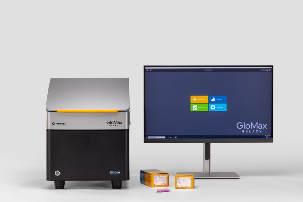
Traditional approaches to decipher cell-to-cell signal rely on complex, time-intensive measures that only approximated the findings acquired through bioluminescence. That’s where the GloMax® Galaxy Bioluminescence Imager comes in. This new tool will enhance your ability to visualize proteins using NanoLuc® technology, going beyond simple numeric outputs to reveal what’s happening in individual cells.

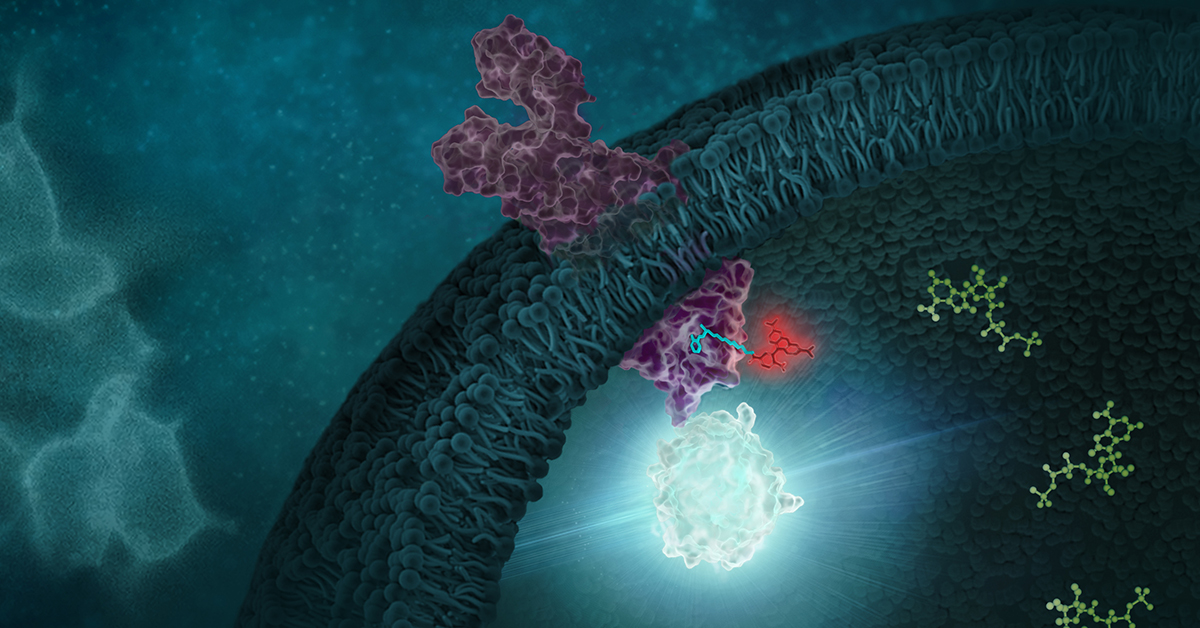
NanoLuc® technology is well-known for its ability to assist in detecting subtle protein interactions in complex biological systems. This bright luminescent enzyme enables a much broader linear range than fluorescence, improving detection of small changes in protein activity, such as proteins interacting. Microplate readers measuring NanoLuc® assays rely on signal generated from many cells. This results in an approximation of what is occurring biologically. Truly validating those luminescent readings within a cell population has been challenging—until now. The GloMax® Galaxy allows you to perform bioluminescence imaging, moving beyond the numbers, offering the power to visualize protein interactions directly.

Continue reading “Visualize Protein:Protein Interactions with Bioluminescence Imaging”

A BiT or BRET, Which is Better?

Now that Promega is expanding its offerings of options for examining live-cell protein interactions or quantitation at endogenous protein expression levels, we in Technical Services are getting the question about which option is better. The answer is, as with many assays… it depends! First let’s talk about what are the NanoBiT and NanoBRET technologies, and then we will provide some similarities and differences to help you choose the assay that best suits your individual needs.

Continue reading “A BiT or BRET, Which is Better?”

For Protein Complementation Assays, Design is Everything

Most, if not all, processes within a cell involve protein-protein interactions, and researchers are always looking for better tools to investigate and monitor these interactions. One such tool is the protein complementation assay (PCA). PCAs use  a reporter, like a luciferase or fluorescent protein, separated into two parts (A and B) that form an active reporter (AB) when brought together. Each part of the split reporter is attached to one of a pair of proteins (X and Y) forming X-A and Y-B. If X and Y interact, A and B are brought together to form the active enzyme (AB), creating a luminescent or fluorescent signal that can be measured. The readout from the PCA assay can help identify conditions or factors that drive the interaction together or apart.

A key consideration when splitting a reporter is to find a site that will allow the two parts to reform into an active enzyme, but not be so strongly attracted to each other that they self-associate and cause a signal, even in the absence of interaction between the primary proteins X and Y. This blog will briefly describe how NanoLuc® Luciferase was separated into large and small fragments (LgBiT and SmBiT) that were individually optimized to create the NanoBiT® Assay and show how the design assists in monitoring protein-protein interactions.

Continue reading “For Protein Complementation Assays, Design is Everything”