What’s In YOUR Protein? Optimizing Protease Digestions to Get the Inside Scoop

It’s time to analyze your protein and you are trying to decide where to begin. You are asking questions like: Which protease do I choose? How much enzyme should I use in my digest? How long should I perform my digest?

Unfortunately, there is no one-size fits all answer to this type of question other than… “well it depends.” All protease digests will be a balance between denaturing the protein sample to allow access to cleavage sites, optimizing conditions for the protease to function, and compatibility with your workflow and downstream applications. We provide general guidelines that work for most samples, but frequently you will need to optimize the conditions need for your specific sample and application.

Here, I use the example of a trypsin digest for downstream mass spectrometry to highlight key questions to ask and factors that can be optimized for any digest.

Continue reading “What’s In YOUR Protein? Optimizing Protease Digestions to Get the Inside Scoop”

Sample preparation: A critical step for consistent protein phosphorylation data

MSextractcroppedProtein phosphorylation is a very important protein post-translational modification that controls many cellular processes including metabolism, transcriptional and translation regulation, degradation of proteins, cellular signaling and communication, proliferation, differentiation, and cell survival (1). Approximately 35% of human proteins are phosphorylated. Phosphoproteins are low in abundance, and, therefore, are challenging to detect and characterize by mass spectrometry. Different enrichment systems have been developed to isolate phosphopeptides. Among these techniques, immobilized metal affinity chromatography (IMAC) using Fe3+ and Ga3+ has been widely used for the enrichment of phosphopeptides.
Typical experimental workflows are tedious and consist of numerous steps, including sample collection and cell lysis. One of the major challenges of the process is to maintain the in vivo phosphorylation state of the proteins throughout the preparation process

To evaluate the effect of sample collection protocols on the global phosphorylation status of the cell, a recent paper by Kashin et al. compared different sample workflows by metabolic labeling and quantitative mass spectrometry on Saccharomyces cerevisiae cell cultures (2).

Three different sample collection workflows were evaluated: two that used denaturating conditions and involved mixing of cell cultures with an excess of either ethanol (EtOH) at −80 °C or trichloroacetic acid (TCA), and a third under nondenaturing conditions and washing the cells in PBS.

Their data suggest that either TCA or EtOH sample collection protocols introduced lower collection bias than the PBS protocol. It was also suggested that similar studies be carried out to determine what effects sample preparation has on other post translation modifications such as acetylation or ubiquitination.

Literature Cited

  1. Thingholm T.E. et al, (2009) Analytical strategies for phosphoproteomics. Proteomics 9,1451–68
  2. Kanshin, E. et al. (2015)  Sample Collection Method Bias Effects in Quantitative Phosphoproteomics. J  Proteome Res. 14, 2998-04.

Purifying HIS-Tagged Proteins from Insect and Mammalian Cells

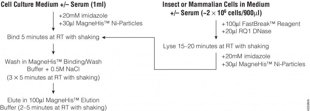
MSextractcroppedMany different polypeptide fusion partners or affinity tags have been developed to facilitate purification of target proteins. The most commonly used tag for the purification and detection of recombinant expressed proteins is the His tag. Cloning vectors designed to generate His-tagged proteins contain 5–10 histidine residues at either the C- or N terminus of the expressed protein. The His tag adds only 0.84kDa to the mass of the protein and is nonimmunogenic. Also, because the tertiary structure of the tag is not important for purification, His-tagged proteins can be purified using native or denaturing conditions. The affinity of histidine residues for immobilized nickel allows selective purification of His-tagged proteins. The MagneHis™ Ni-Particles can bind up to 1mg of His-tagged protein per milliliter of particles providing a fast, efficient method for purifying His-tagged proteins with high yield and low background in a highly scalable format.

Bacterial expression of recombinant His-tagged proteins is a common technique. However, use of other systems, such as Sf9 insect cells,or HeLa or CHO mammalian cells for expression of recombinant proteins either intracellularly or secreted into the culture medium is increasing. These eukaryotic expression systems may allow more natural processing and modification of recombinant His-tagged proteins.
The following article:  illustrates the use of FastBreak™ Cell Lysis Reagent and the MagneHis™ Protein Purification System with insect and mammalian cell lysates. Proteins are purified from culture medium in the presence or absence of serum with only minior modifications to the standard protocol for bacterial cultures are required for purification from these diverse sources.
4550MA-(1)


His-Tagged Fusion Proteins: Application Update

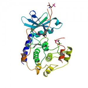
Crystal structure of Polyhistidine tagged recombinant catalytic subunit of cAMP-dependent protein kinase. Credit: StructureID=1fmo; DOI=http://dx.doi.org/10.2210/pdb1fmo/pdb;

Researchers often need to purify a single protein for further study. One method for isolating a specific protein is the use of affinity tags. Affinity purification tags can be fused to any recombinant protein of interest, allowing fast and easy purification following a procedure that is based on the affinity properties of the tag.

The most commonly used tag to purify and detect recombinant expressed proteins is the polyhistidine tag. Protein purification using polyhistidine tags relies on the affinity of histidine residues for immobilized metal such as nickel, which allows selective protein purification. The metal is immobilized to a support through complex formation with a chelate that is covalently attached to the support.

Polyhistidine tags offer several advantages for protein purification. The small size of the polyhistidine tag renders it less immunogenic than other larger tags. Therefore, the tag usually does not need to be removed for downstream applications following purification.

A large number of commercial expression vectors that contain polyhistidine are available. The polyhistidine tag may be placed on either the N- or C-terminus of the protein of interest.

And finally, the interaction of the polyhistidine tag with the metal does not depend on the tertiary structure of the tag, making it possible to purify otherwise insoluble proteins using denaturing conditions. The resulting purified protein can be used for a variety of applications.

The following references illustrate examples of some of the most common post purification applications with fusion proteins containing a polyhistidine tag:

Enzymatic assays

  1. Negi, V-S. et al. (2014) A carbon nitrogen Lyase from Leucaena leucocephala catalyzes the first Step of mimosine degradationPlant Physiol. 164,  922–34.
  2. Yu, S. et al. (2013) Syk Inhibits the activity of protein kinase A by phosphorylation tyrosine of the catalytic subunitJ. Biol. Chem. 288, 10870-81.
  3. Rusconi, B. et al. (2013) Discovery of catalases in members of the Chiamydiales order. J. Bact. 195, 3543–51.

Structural analysis

  1. Araiso, Y. et al. (2014) Crystal structure of Saccharomyces cerevisiae mitochondrial GatFAB a novel subunit assembly in tRNA –dependent amidotransferases. Nucl.Acids. Res.(available only online).
  2. Someya, T. et al. (2012) Crystal Structure of Hfq from Bacillus subtilis in complex with SELEX-dervived RNA aptamer: insight into RNA-binding properties of bacterial Hfq. Nucl. Acid Res. 40, 1856-67.

Protein pulldowns

  1. Yun, S-C. et al. (2010) Pmr a Histone-like protein H1 (H-NS) family protein encoded by the IncP-7 plasmid pCAR1, is a key global regulator that alters host functionJ.Bact. 192, 4720–31.
  2. Haim, H. et al. (2010)  Cytokeratin 8 interacts with clumping factor B: a new possible virulence factor target. Microbiology 156, 3710-21.

Additional Resources
His-tagged Protein Purification Systems

Purification of biotinylated proteins

Biotinylation is an attractive approach for protein complex purification due to the very high affinity (Kd = 10–15 M) of avidin/streptavidin for biotinylated templates. With typical avidin or streptavidin, the biotin-binding affinity is so great that purification with these traditional media require denaturing conditions for elution,such as 8 M Guanidine•HCl at pH 1.5 or boiling in reducing SDS-PAGE sample loading buffer. To avoid these harsh conditions SoftLink™ Soft Release Avidin resin can be used. These particles consist of monomeric avidin coupled to a methylacrylate resin.

This resin provides the same specificity of binding to biotin afforded by tetrameric biotin, but enables the release of biotinlylated molecules under mild nondenaturing conditions (5mM biotin).

The following are recent references that used the SoftLink™ Resin for the noted application:

Kashwayama, Y. et al. (2010) Identification of a substrate-binding site in a peroxisomal beta-oxidation enzyme by photoaffinity labeling with a novel palmitoyl derivative. J. Biol. Chem. 285, 26315–25. (Purification of photoaffinity labeled proteins for subquenant binding/activity experiments)

Takahashi, M. et al. (2010) Tailor-made RNAi knockdown against triplet repeat disease-causing alleles. Proc. Natl. Acad. Sci. 107, 21731-36 (Innovated procedure using biotin labeled cDNAs for the identification of nucleotide variations)

Kress, D. et al. (2009) An asymmetric model for Na+-translocating glutaconyl-CoA decarboxylases. J. Biol.Chem. 284, 28401–9 (Purification of Clostridium biotin carrier proteins that play a role in decarboxylation)

Akahori, Y. et al. (2009) Characterization of neutralizing epitopes of varicella-zoster virus glycoprotein H. J. Virol. 83, 2020–4. (purification of double stranded cDNA fragments amplified by PCR with a biotin-tagged PCR primer)

Shonsey, E.M. et al. (2008) Inactivation of human liver bile acid CoA:amino acid N-acyltransferase by the electrophilic lipid, 4-hydroxynonenal. J. Lipid Res. 49, 282–94 (purification of recombinant protein expressed in E.coli containing C-terminal avidin tag)

Andachi, Y. et al. (2008) A novel biochemical method to identify target genes of individual microRNAs: identification of a new Caenorhabditis elegans let-7 target. RNA 14, 2440–51 (purification of double stranded cDNA fragments amplified by PCR with a biotin-tagged PCR primer)

For more information about SoftLink™ Soft Release Avidin Resin, please visit our website.

6X His Protein Pulldowns: An Alternative to GST

ResearchBlogging.orgPull-down assays probe interactions between a protein of interest that is expressed as fusion protein (e.g.,
(e.g., bait) and the potential interacting partners (prey).

In a pull-down assay one protein partner is expressed as a fusion protein (e.g., bait protein) in E. coli and then immobilized using an affinity ligand specific for the fusion tag. The immobilized
bait protein can then be incubated with the prey protein. The source of the prey protein depends on whether the experiment is designed to confirm an interaction or to identify new interactions. After a series of wash steps, the entire complex can be eluted from the affinity support using SDS-PAGE loading buffer or by competitive analyte elution, then evaluated by SDS-PAGE.

Successful interactions can be detected by Western blotting with specific antibodies to both the prey and bait proteins, or measurement of radioactivity from a [35S] prey protein. bait) and potential interacting partners (prey).

The most commonly used method to generate a bait protein is expression as a fusion protein contain a GST (glutathione-S transferase) tag in E. coli. This is followed by immobilization on particles that contain reduced glutathione, which binds to the GST tag of the fusion protein. The primary advantage of a GST tag is that it can increase the solubility of insoluble or semi-soluble proteins expressed in E. coli.

Among fusion tags, His-tag is the most widely used and has several advantages including: 1) It’s small in size, which renders it less immunogenically active, and often it does not need to be removed from the purified protein for downstream applications; 2) There are a large number of commercial vectors available for expressing His-tagged proteins; 3) The tag may be placed at either the N or C terminus; 4) The interaction of the His-tag does not depend on the tag structure, making it possible to purify otherwise insoluble proteins using denaturing conditions. Continue reading “6X His Protein Pulldowns: An Alternative to GST”