NanoBRET™ Assays to Analyze Virus:Host Protein:Protein Interactions in Detail

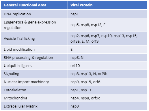
Recently, Gordon et al. published an atlas of protein:protein interactions of all proposed SARS-CoV-2 proteins expressed individually in HEK 293 cells (Table 1). The study tagged each of the viral proteins with an epitope tag and performed a pull-down of the expressed protein followed by trypsin digestion and mass spec analysis, a process referred to as affinity purification–mass spec analysis. The group identified 332 human proteins interacting with 27 SARS-CoV-2 proteins.

The interactions identified in the HEK 293 cells helped Appelberg et al. analyze interactions over time in SARS-CoV-2-infected Huh7 cells. Gordon et al. used the PPI data to identify FDA-approved drugs, drugs in clinical trials, and pre-clinical compounds that bound to the identified human proteins and labs in New York and Paris tested some of these drugs for antiviral effects.   

Table 1. The general functional area of human proteins identified to interact with individually expressed SARS-CoV-2 proteins as reported by Gordon, et al. (1). The SARS-CoV-2 proteins are classified as non-structural proteins (nsp#), structural proteins (E, M, and N) and accessory proteins (orf#).  
Continue reading “NanoBRET™ Assays to Analyze Virus:Host Protein:Protein Interactions in Detail”

Research Teams Demonstrate Bivalent Binding of a Novel Bromodomain Protein Inhibitor

13305818-cr-da-nanoluc-application_ligund

Today’s blog is written by guest blogger Kristin Huwiler from our Cellular Analysis and Proteomics Group.

Two research collaborations, one in Europe and a second in the US, have just published in Nature Chemical Biology (1,2) on the identification of BET inhibitors (bi-BETs) that bind via a bivalent mechanism to both bromodomains of BRD4. These bivalent chemical inhibitors exhibit high cellular potency and affinity relative to their monovalent predecessors. By developing high-affinity ligands that engage both bromodomains simultaneously within BRD4, the authors illustrate a concept that may be applicable in the development of selective, potent ligands for other multi-domain proteins. Here we review the work presented in the Waring et al. paper using the Promega NanoBRET™ Technologies to characterize the mechanism of action of their bivalent probe.

The bromodomain and extraterminal (BET) sub-family are some of the most studied bromodomain-containing proteins (3). The BET subfamily of proteins contain two separate bromodomains. BRD4 is one well studied member of the BET sub-family. Several small molecule inhibitors that target BRD4 have been developed as potential therapeutics for various cancers with promising initial studies, but to date are all monovalent, binding each bromodomain of the BET family members separately (2).

Continue reading “Research Teams Demonstrate Bivalent Binding of a Novel Bromodomain Protein Inhibitor”

NanoBiT™ Assay: Transformational Technology for Studying Protein Interactions Named a Top 10 Innovation of 2015

21416943-wb-cr-uw-nanobit-pm-page-hero
For three out of the last four years, we have been honored to have one of our key technologies named a Top 10 Innovation by The Scientist. This year the innovative NanoBiT™ Assay (NanoLuc® Binary Technology) received the recognition. NanoBiT™ is a structural complementation reporter based on NanoLuc® Luciferase, a small, bright luciferase derived from the deep sea shrimp Oplophorus gracilirostris.

Using plasmids that encode the NanoBiT complementation reporter, you can make fusion proteins to “report” on protein interactions that you are studying. One of the target proteins is fused to the 18kDa subunit; the other to the 11 amino acid subunit. The NanoBiT™ subunits are stable, exhibiting low self-affinity, but produce an ultra-bright signal upon association. So, if your target proteins interact, the two subunits are brought close enough to each other to associate and produce a luminescent signal. The strong signal and low background associated with a luminescent system, and the small size of the complementation reporter, all help the NanoBiT™ assay overcome the limitations associated with traditional methods for studying protein interactions.

The small size reduces the chances of steric interference with protein interactions. The ultra bright signal, means that even interactions among proteins present in very low amounts can be detected and quantified–without over-expressing large quantities of non-native fusion proteins and potentially disrupting the normal cellular environment. And the NanoBiT™ assay can be performed in real time, in live cells.

The NanoBiT™ assay is already being deployed in laboratories to help advance understanding of fundamental cell biology. You can see how one researcher is already taking full advantage of this innovative technology in the video embedded below:

Visit the Promega web site to see more examples more examples how the NanoBiT™ assay can break through the traditional limitations for studying protein interactions in cells.

You can read the Top 10 article in The Scientist here.

Interrogating Protein Interactions: An Infographic for NanoBRET™ Assay Design

Yesterday my fellow blogger, Kari, posted a review of the ACS Chemical Biology paper describing a new BRET platform for analyzing protein-protein interactions. If you are interested in studying induction and inhibition of protein interactions in real time, take a look at the infographic below to learn how to develop a NanoBRET™ Assay to monitor your protein of interest.

21421668-IG-CR-NanoBRET-Infographic-Final

If We Could But Peek Inside the Cell …Quantifying, Characterizing and Visualizing Protein:Protein  Interactions

14231183 WB MS Protein Interactions Hero Image 600x214

Robert Hooke first coined the term “cell” after observing  plant cell walls through a light microscope—little empty chambers, fixed in time and space. However,  cells are anything but fixed.

Cells are dynamic: continually responding to a shifting context of time, environment, and signals from within and without. Interactions between the macromolecules within cells, including proteins, are ever changing—with complexes forming, breaking up, and reforming in new ways. These interactions provide a temporal and special framework for the work of the cell, controlling gene expression, protein production, growth, cell division and cell death.

Visualizing and measuring protein:protein interactions at the level of the cell without perturbing them is the goal of every cell biologist.

A recent article by Thomas Machleidt et al. published in ACS Chemical Biology, describes a new technology that brings us closer to being able to realize that goal.

Continue reading “If We Could But Peek Inside the Cell …Quantifying, Characterizing and Visualizing Protein:Protein  Interactions”