Better NGS Size Selection

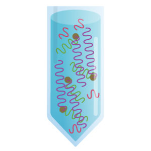
One of the most critical parts of a Next Generation Sequencing (NGS) workflow is library preparation and nearly all NGS library preparation methods use some type of size-selective purification. This process involves removing unwanted fragment sizes that will interfere with downstream library preparation steps, sequencing or analysis.

Different applications may involve removing undesired enzymes and buffers or removal of nucleotides, primers and adapters for NGS library or PCR sample cleanup. In dual size selection methods, large and small DNA fragments are removed to ensure optimal library sizing prior to final sequencing. In all cases, accurate size selection is key to obtaining optimal downstream performance and NGS sequencing results.

Current methods and chemistries for the purposes listed above have been in use for several years; however, they are utilized at the cost of performance and ease-of-use. Many library preparation methods involve serial purifications which can result in a loss of DNA. Current methods can result in as much as 20-30% loss with each purification step. Ultimately this may necessitate greater starting material, which may not be possible with limited, precious samples, or the incorporation of more PCR cycles which can result in sequencing bias. Sample-to-sample reproducibility is a daily challenge that is also regularly cited as an area for improvement in size-selection.

Continue reading “Better NGS Size Selection”

How Do I Choose the Right GoTaq® Product to Suit My Needs for EndPoint PCR?

We offer a wide array of GoTaq® DNA Polymerases, Buffers and Master Mixes, so we frequently answer questions about which product would best suit a researcher’s needs. On the Taq Polymerase Page, you can filter the products by clicking the categories on the left hand side of the page to narrow down your search. Here are some guidelines to help you select the match that will best suit your PCR application. Continue reading “How Do I Choose the Right GoTaq® Product to Suit My Needs for EndPoint PCR?”

T-Vector Cloning: Answers to Frequently Asked Questions

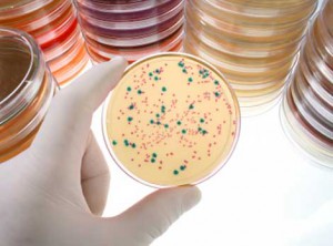
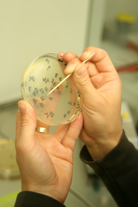
Blue/White colony screening helps you pick only the colonies that have your insert.

Q: Can PCR products generated with GoTaq® DNA Polymerase be used to for T- vector cloning?

A: Yes. GoTaq® DNA Polymerase is a robust formulation of unmodified Taq Polymerase. GoTaq®DNA Polymerase lacks 3’ →5’ exonuclease activity (proof reading) and also displays non-template–dependent terminal transferase activity that adds a 3′ deoxyadenosine (dA) to product ends. As a result, PCR products amplified using GoTaq® DNA Polymerase will contain A-overhangs which makes it suitable for T-vector cloning.

We have successfully cloned PCR products generated using GoTaq® and GoTaq® Flexi DNA Polymerases into the pGEM®-T (Cat.# A3600), pGEM®-T Easy (Cat.# A1360) and pTARGET™ (Cat.# A1410) Vectors.

Q: Can GoTaq® Long PCR Master Mix be used for T-Vector Cloning?

A: Yes it can. GoTaq® Long PCR Master Mix utilizes recombinant Taq DNA polymerase as well as a small amount of a recombinant proofreading DNA polymerase. This 3´→5´ exonuclease activity (proof reading) enables amplification of long targets. Despite the presence of a small amount of 3´→5´ exonuclease activity, the GoTaq® Long PCR Master Mix generates PCR products that can be successfully ligated into the pGEM®-T Easy Vector System.

We have demonstrated that GoTaq® Long PCR Master Mix successfully generated DNA fragments that could be ligated into pGEM®-T Easy Vector System without an A-tailing procedure, and with ligation efficiencies similar to those observed with the GoTaq® Green Master Mix.

For details refer to Truman, A., Hook, B. and Wieczorek, D. Using GoTaq® Long PCR Master Mix for T-Vector Cloning.

Tip: For cloning blunt-ended PCR fragments into T-vectors, use the A-tailing protocol discussed in the pGEM®-T and pGEM®-T Easy Technical Manual #TM042.

Q: How do I prepare PCR products for ligation? What products can be used to purify the DNA?

Continue reading “T-Vector Cloning: Answers to Frequently Asked Questions”

Dealing with PCR Inhibitors

Inhibition

The polymerase chain reaction (PCR) has revolutionized modern biology as a quick and easy way to generate amazing amounts of genomic data. However, when PCR doesn’t work, it can be frustrating. At these times, PCR and reverse transcription PCR (RT-PCR) inhibitors seem to be everywhere: They lie dormant in your starting material and can co-purify with the template of interest, and they can be introduced during sample handling or reaction setup. The effects of these inhibitors can range from partial inhibition and underestimation of the target nucleic acid amount to complete amplification failure. What is a scientist to do?

Continue reading “Dealing with PCR Inhibitors”

Top Ten Tips for Successful PCR

We decided to revisit a popular blog from our Promega Connections past for those of you in the amplification world. Enjoy:

magnesium-31

    • Modify reaction buffer composition to adjust pH and salt concentration.
    • Titrate the amount of DNA polymerase.
    • Add PCR enhancers such as BSA, betaine, DMSO, nonionic detergents, formamide or (NH4)2SO4.
    • Switch to hot-start PCR.
    • Optimize cycle number and cycling parameters, including denaturation and extension times.
    • Choose PCR primer sequences wisely.
    • Determine optimal DNA template quantity.
    • Clean up your DNA template to remove PCR inhibitors.
    • Determine the optimal annealing temperature of your PCR primer pair.

[Drum roll please]…and the  most important thing you can do to improve your PCR results is:

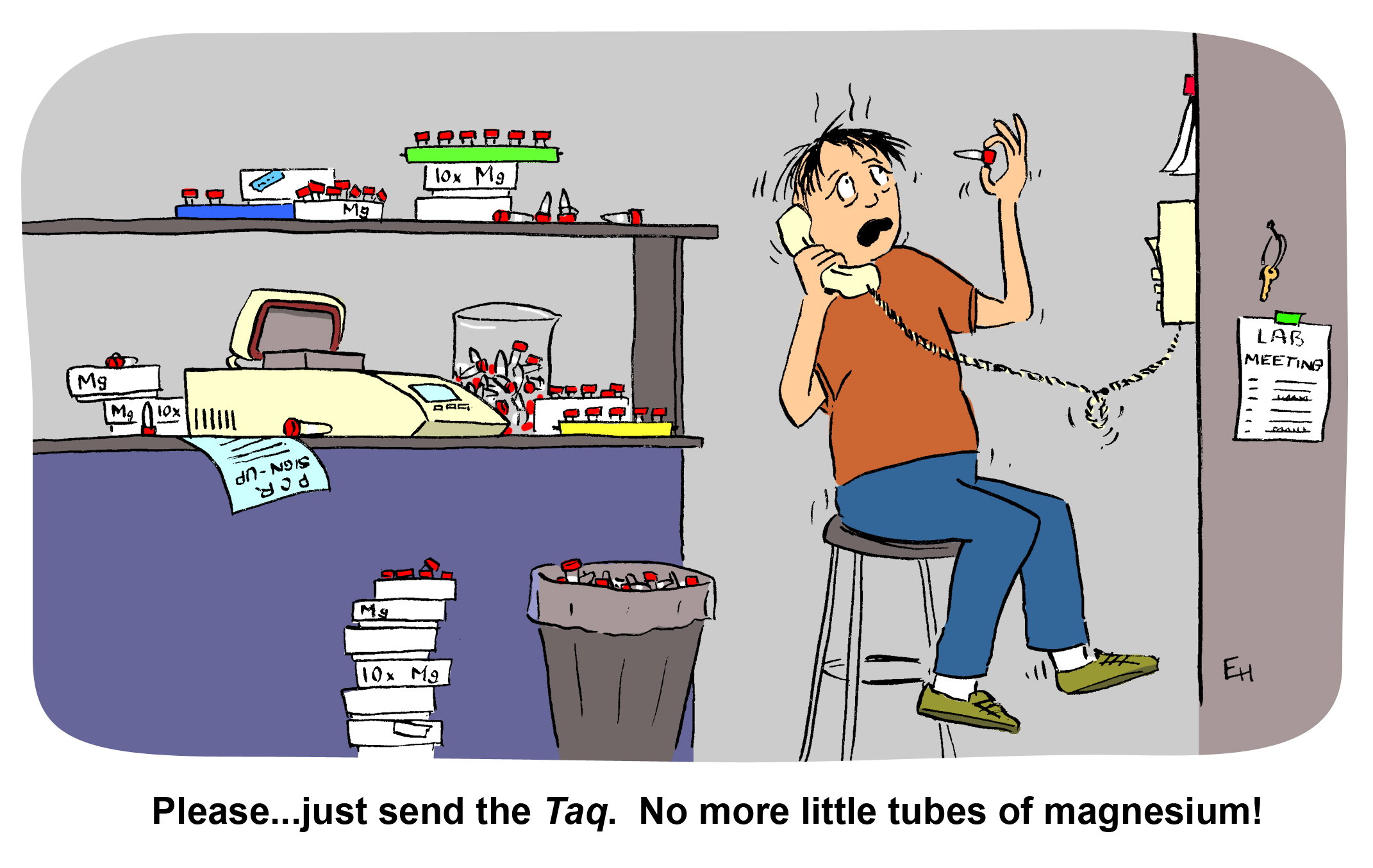
  • Titrate the magnesium concentration.

DNA Purification, Quantitation and Analysis Explained

WebinarsYesterday I listened in on the Webinar “Getting the Most Out of Your DNA Analysis from Purification to Downstream Assays”, presented by Eric Vincent–a Product Manager in the Promega Genomics group.

This is the webinar for you if you have ever wondered about the relative advantages and disadvantages of the many methods available for DNA purification, quantitation and analysis, or if you are comparing options for low- to high-throughput DNA purification. Eric presents a clear analyses of each of the steps in a basic DNA workflow: Purification, Quantitation, Quality Determination, and Downstream Analysis, providing key considerations and detailing the potential limitations of the methods commonly used at each step.

The DNA purification method chosen has an affect on the quality and integrity of the DNA isolated, and can therefore affect performance in downstream assays. Accuracy of quantitation also affects success, and the various downstream assays themselves (such as end-point PCR, qPCR, and sequencing) each have different sensitivities to factors such as DNA yield, quality, and integrity, and the presence of inhibitors. Continue reading “DNA Purification, Quantitation and Analysis Explained”

Troubleshooting T-Vector Cloning

Why do few of my pGEM®-T or pGEM®-T Easy Vector clones contain the PCR product of interest?

There are several possible reasons why the PCR product may not be recovered after ligation, bacterial transformation and plating when using the pGEM®-T or pGEM®-T Easy Vector Systems.

The PCR fragment may not be A-tailed. Without the A overhangs, the PCR product cannot be ligated into a T vector. Use a nonproofreading DNA polymerase like GoTaq® DNA Polymerase for PCR. If a proofreading DNA polymerase is used, A overhangs will need to be added. Purify the PCR fragment, and set up an A-tailing reaction (see the pGEM®-T and pGEM®-T Easy Vector Systems Technical Manual #TM042). The A-tailed product can be added directly to the ligation as described in the pGEM®-T or pGEM®-T Easy Vector protocol.

The insert:vector ratio may not be optimal. The ideal ratio for each insert to a vector can vary. For example, the Control Insert DNA works well at a 1:1 ratio, but another insert may be ligated more efficiently at a 3:1 ratio. Check the integrity and quantity of your PCR fragment by gel analysis. Optimize the insert:vector ratio (see Technical Manual #TM042).

Multiple PCR products were amplified and cloned into the pGEM®-T or pGEM®-T Easy Vector. Other amplification products including primer dimers will compete for ligation into the T vector, decreasing the possibility that the desired insert will be cloned. To minimize other competing products, gel purify the PCR fragment of interest.

Promega Technical Services Scientists are here to assist you in troubleshooting your experiments at any time. Contact Technical Services.

Related Posts

PCR-Based Diagnosis Wins by a Mile In the White-Nose Syndrome Race

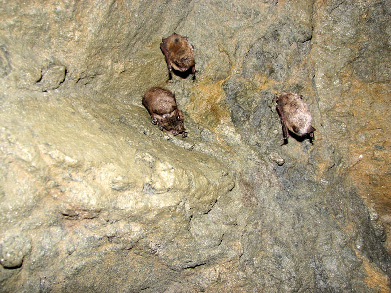
Affected bats in a cave in MA. Image courtesy of the U.S. Geological Survey.

Since the first photograph of bats with white muzzles in Albany, NY, was published, hibernating bat populations in the northeastern U.S. have been devastated by an emerging disease, White-Nose Syndrome (WNS), which continues to spread throughout the United States and now has been found in two Canadian provinces. Bats suffering from WNS are emaciated with little or no body fat and have a characteristic white fungal growth on their wing membranes, ears and muzzles. Instead of hibernating all winter, these bats can be seen active in the snow, when there is virtually no food available.

The infectious agent that responsible for WNS is a new species of the cold-loving fungus, Pseudogymnoascus destructans (formerly, Geomyces destructans) . Currently, WNS is confirmed either through histological analysis or by fungal culture. Both of these techniques have significant limitations. First, they have turnaround times of at least one week. Secondly, they require large amounts of tissue sample from affected bats, more than can be reasonably taken from live bats. Histological analysis is a laborious process that requires highly skilled and trained personnel. Fungal culture can be difficult because bats harbor many bacteria and fungi, and getting a pure culture of a causative organism is not simple. Furthermore, researchers need a quick method for assessing spread of the disease that can provide results quickly.

Polymerase Chain Reaction (PCR), already used for a host of diagnostic tests in humans, plants and animals, is a logical choice. In a paper published in the Journal of Veterinary Diagnostic Investigation, Lorch and colleagues design and evaluate a PCR-based diagnostic method for WNS. They compare the PCR method to a fungal culture method and the “gold standard” traditional histological analysis. Continue reading “PCR-Based Diagnosis Wins by a Mile In the White-Nose Syndrome Race”

Humorous New Types of PCR

Undoubtedly, the polymerase chain reaction (PCR) has revolutionized biological research and has become one of the most common techniques in today’s laboratory. At times, it seems that a new variation of PCR is described in the literature every month. You might think that you are familiar with the dozens of PCR variations, but I am guessing that you haven’t heard of some of these.

Continue reading “Humorous New Types of PCR”