A Diamond in the Rough: New Applications of Diamond Nucleic Acid Dye

Diamond™ Nucleic Acid Dye (Cat# H1181) is a safe, inexpensive and sensitive fluorescent dye option that binds to single-stranded and double-stranded DNA and RNA. Diamond™ Dye typically is used for staining electrophoresis gels to visualize nucleic acids in a similar to its carcinogenic counterpart, ethidium bromide. However Diamond™ Dye has several advantages: gels stained with Diamond™ Dye can be visualized using either UV or blue-light transilluminators. Also, a wash step after staining is not necessary when using Diamond™ Dye, unlike what is typically recommended for ethidium bromide.

Besides staining electrophoresis gels, there are other applications for this diamond in the rough. Highlighted below are two fascinating uses of this multifaceted tool: touch DNA localization and qPCR detection.

Continue reading “A Diamond in the Rough: New Applications of Diamond Nucleic Acid Dye”

Fluorescence Dye-Based Quantitation: Sensitive and Specific for NGS Applications

This is the third post in a series of blogs on quantitation for NGS applications written by guest blogger Adam Blatter, Product Specialist in Integrated Solutions at Promega.

Fluorescent dye-based quantitation uses specially designed DNA binding compounds that intercalate only with double stranded DNA molecules. When excited by a specific wavelength of light, only dye in the DNA-bound state will fluoresce. These aspects of the technique contribute to low background signal, and therefore the ability to accurately and specifically detect very low quantities of DNA in solution, even the nanogram quantities used in NGS applications.

For commercial NGS systems, such as the Nextera Rapid Capture Enrichment Protocol by Illumina, this specificity and sensitivity of quantitation are critical. The Nextera protocol is optimized for 50ng of total genomic DNA. A higher mass input of genomic DNA can result in incomplete tagmentation, and larger insert sizes, which can adversely affect enrichment. A lower mass input of genomic DNA or low-quality DNA can generate smaller than expected inserts, which can be lost during subsequent cleanup steps, giving lower diversity of inserts.

Continue reading “Fluorescence Dye-Based Quantitation: Sensitive and Specific for NGS Applications”

Nucleic Acid Quantitation by UV Absorbance: Not for NGS

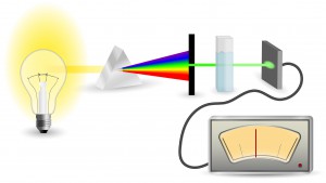
schematic diagram of UV-Vis Absorbance Method
For UV-Vis Spectrophotometry, light is split into its component wavelengths and directed through a solution. Molecules in the solution absorb specific wavelengths of light.

This is the second in a series of four blogs about Quantitation for NGS is written by guest blogger Adam Blatter, Product Specialist in Integrated Solutions at Promega.

Perhaps the most ubiquitous quantitation method is UV-spectrophotometry (also called absorbance spectroscopy). This technique takes advantage of the Beer-Lambert Law: an observation that many compounds absorb UV-Visible light at unique wavelengths, and that for a fixed path length the absorbance of a solution is directly proportional to the concentration of the absorbing species. DNA, for example has a peak absorbance at 260nm (A260nm).

This method is user friendly, quick and easy. But, it has significant limitations, especially when quantitating samples for NGS applications.

Continue reading “Nucleic Acid Quantitation by UV Absorbance: Not for NGS”

When Every Step Counts: Quantitation for NGS

13170MA-800x277This series of blogs about Quantitation for NGS is written by guest blogger Adam Blatter, Product Specialist in Integrated Solutions at Promega.

As sequencing technology races toward ever cheaper, faster and more accurate ways to read entire genomes, we find ourselves able to study biological systems at a level never before possible. From basic science to translational research, massively parallel sequencing (also known as next-generation sequencing or NGS) has opened up new avenues of inquiry in genomics, oncology and ecology.

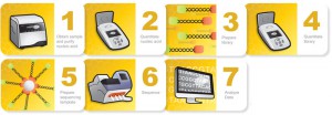
Many commercial sequencing platforms have been established (e.g., Illumina, IonTorrent, 454, PacBio), and new technologies are developed every day to enable new and unique applications. However, all of these platforms and technologies work off the same general principle: nucleic acid must be extracted from a sample, arranged into platform-specific library constructs, and loaded into the sequencer. Regardless of the sample type or the platform used, every step throughout this workflow is critical for successful results. An often overlooked part of the NGS workflow is sample quantitation. Here we are presenting the first in a series of four short blogs about the critical step of quantitation in NGS workflows.

Sample input is critical to NGS in terms of both quality and quantity. Knowing how much DNA you have, often in nanogram quantities, can make the difference between success and failure. There are several key points in the NGS workflow where sample quantitation is important before you can proceed:

  • After initial nucleic acid extraction from the sample matrix and before proceeding with library preparation
  • Post-library preparation when pooling barcoded libraries for multiplexing
  • Final pooled library quantitation immediately before loading for sequencing

There are several common methods for quantitating nucleic acids: UV-spectroscopy, Fluorescence spectoscopy, real-time quantitative PCR (qPCR). Because of inherent differences in sensitivity, specificity, time and cost, each of these techniques pose certain advantages and disadvantages with respect to the specific sample you are quantitating. Our next three blogs will discuss each of these methods against the backdrop of quantitating samples for NGS applications.


Read Part 2: Nucleic Acid Quantitation by UV Absorbance: Not for NGS

Read Part 3: Fluorescence Dye-Based Quantitation: Sensitive and Specific for NGS Applications

Read Part 4: Real-Time (Quantitative) qPCR for Quantitating Library Prep before NGS