Revealing Time of Death: The Microbiome Edition

Forensic analysts have long sought precision when determining time of death. While on crime scene investigation television shows, the presence of insects always seems to reveal when a person died, there are many elements to account for, and the probable date may still not be accurate. Insects arrive days after death if at all (e.g., if the body is found indoors or after burial), and the stage of insect activity is influenced by temperature, weather conditions, seasonal variation, geographic location and other factors. All this makes it difficult to estimate the postmortem interval (PMI) of a body discovered an unknown time after death. One way to make estimating PMI less subjective would be to have calibrated molecular markers that are easy to sample and are not altered by environmental variabilities.

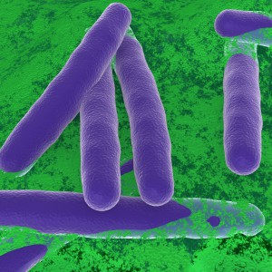
Bacterial communities called microbiomes have been frequently in the news. The influence of these microbes encompass living creatures and the environment. Not surprisingly, research has focused on the influence of microbiomes on humans. For example, changes in gut microbiome seem to affect human health. Intriguingly, microbiomes may also be a key to determining time of death. The National Institute of Justice (NIJ) has funded several projects focused on the forensic applications of microbiomes. One focus involves the necrobiome, the community of organisms found on or around decomposing remains. These microbes could be used as an indicator of PMI when investigating human remains. Recent research published in PLOS ONE examined the bacterial communities found in human ears and noses after death and how they changed over time. The researchers were interested in developing an algorithm using the data they collected to estimate of time of death.

Continue reading “Revealing Time of Death: The Microbiome Edition”
Friday Cartoon Fun: Take My Samples, Please!

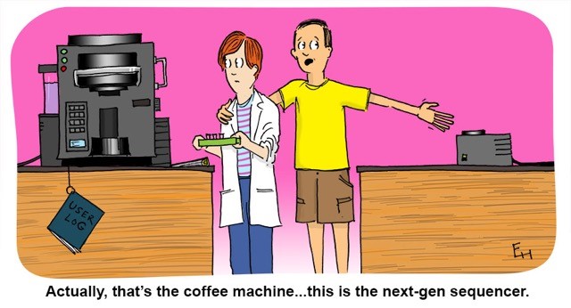
Instruments can make our lives easier in the lab. Place your samples inside an instrument and let it do all the work—isolating nucleic acids or reading and analyzing a multiwell plate—while you walk away to read a new research paper or prepare for the next step in your experiment. However, with the array of machines now available to scientists worldwide, some confusion may result in the laboratory. Has this ever happened to you?

eh_March2016
Copyright Ed Himelblau

Real-Time (quantitative) qPCR for Quantitating Library Prep before NGS

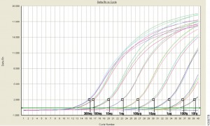
Real-Time (or quantitative, qPCR) monitors PCR amplification as it happens and allows you to measure starting material in your reaction.

This the last in a series of four blogs about Quantitation for NGS is written by guest blogger Adam Blatter, Product Specialist in Integrated Solutions at Promega.

When it comes to nucleic acid quantitation, real-time or quantitative (qPCR) is considered the gold standard because of its unmatched performance in senstivity, specificity and accuracy. qPCR relies on thermal cycling, consisting of repeated cycles of heating an cooling for DNA melting and enzyamtic replication. Detection instrumentation is capable of measuring the accumulation of DNA product after each round of amplification in real time.

Because PCR amplifies specific regions of DNA, the method is highly sensitive, specific to DNA, and it can determine whether a sample is truly able to be amplified. Degraded DNA or free nucleotides, which might otherwise skew your quantiation, will not contribute to the signal, and your measurement will be more accurate.

However, while qPCR does provide technical advantages, the method requires special instrumentation, specialized reagents and is a more time-consuming process. In addition, you will probably need to optimize your qPCR assay for each of your targets to achieve your desired results.

Because of the added complexity and cost, qPCR is a technique suited for post-library quantitation when you need to know the exact amount of amplifiable, adapter-ligated DNA.  PCR is the only method capable of specifically targeting these library constructs over other DNA that may be present. This specificity is important because accurate normalization is especially critical for producing even coverage in multiplex experiments where equimolar amounts of several libraries are added to a pooled sample. This normalization process is essential  if your are screening for rare variants that might be lost in background and go undetected if underrepresented in a mixed pool.


Read Part 1: When Every Step Counts: Quantitation for NGS

Read Part 2: Nucleic Acid Quantitation by UV Absorbance: Not for NGS

Read Part 3: Fluorescence Dye-Based Quantitation: Sensitive and Specific for NGS Applications

Fluorescence Dye-Based Quantitation: Sensitive and Specific for NGS Applications

This is the third post in a series of blogs on quantitation for NGS applications written by guest blogger Adam Blatter, Product Specialist in Integrated Solutions at Promega.

Fluorescent dye-based quantitation uses specially designed DNA binding compounds that intercalate only with double stranded DNA molecules. When excited by a specific wavelength of light, only dye in the DNA-bound state will fluoresce. These aspects of the technique contribute to low background signal, and therefore the ability to accurately and specifically detect very low quantities of DNA in solution, even the nanogram quantities used in NGS applications.

For commercial NGS systems, such as the Nextera Rapid Capture Enrichment Protocol by Illumina, this specificity and sensitivity of quantitation are critical. The Nextera protocol is optimized for 50ng of total genomic DNA. A higher mass input of genomic DNA can result in incomplete tagmentation, and larger insert sizes, which can adversely affect enrichment. A lower mass input of genomic DNA or low-quality DNA can generate smaller than expected inserts, which can be lost during subsequent cleanup steps, giving lower diversity of inserts.

Continue reading “Fluorescence Dye-Based Quantitation: Sensitive and Specific for NGS Applications”

Nucleic Acid Quantitation by UV Absorbance: Not for NGS

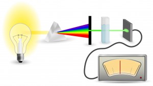
schematic diagram of UV-Vis Absorbance Method
For UV-Vis Spectrophotometry, light is split into its component wavelengths and directed through a solution. Molecules in the solution absorb specific wavelengths of light.

This is the second in a series of four blogs about Quantitation for NGS is written by guest blogger Adam Blatter, Product Specialist in Integrated Solutions at Promega.

Perhaps the most ubiquitous quantitation method is UV-spectrophotometry (also called absorbance spectroscopy). This technique takes advantage of the Beer-Lambert Law: an observation that many compounds absorb UV-Visible light at unique wavelengths, and that for a fixed path length the absorbance of a solution is directly proportional to the concentration of the absorbing species. DNA, for example has a peak absorbance at 260nm (A260nm).

This method is user friendly, quick and easy. But, it has significant limitations, especially when quantitating samples for NGS applications.

Continue reading “Nucleic Acid Quantitation by UV Absorbance: Not for NGS”

When Every Step Counts: Quantitation for NGS

13170MA-800x277This series of blogs about Quantitation for NGS is written by guest blogger Adam Blatter, Product Specialist in Integrated Solutions at Promega.

As sequencing technology races toward ever cheaper, faster and more accurate ways to read entire genomes, we find ourselves able to study biological systems at a level never before possible. From basic science to translational research, massively parallel sequencing (also known as next-generation sequencing or NGS) has opened up new avenues of inquiry in genomics, oncology and ecology.

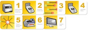
Many commercial sequencing platforms have been established (e.g., Illumina, IonTorrent, 454, PacBio), and new technologies are developed every day to enable new and unique applications. However, all of these platforms and technologies work off the same general principle: nucleic acid must be extracted from a sample, arranged into platform-specific library constructs, and loaded into the sequencer. Regardless of the sample type or the platform used, every step throughout this workflow is critical for successful results. An often overlooked part of the NGS workflow is sample quantitation. Here we are presenting the first in a series of four short blogs about the critical step of quantitation in NGS workflows.

Sample input is critical to NGS in terms of both quality and quantity. Knowing how much DNA you have, often in nanogram quantities, can make the difference between success and failure. There are several key points in the NGS workflow where sample quantitation is important before you can proceed:

  • After initial nucleic acid extraction from the sample matrix and before proceeding with library preparation
  • Post-library preparation when pooling barcoded libraries for multiplexing
  • Final pooled library quantitation immediately before loading for sequencing

There are several common methods for quantitating nucleic acids: UV-spectroscopy, Fluorescence spectoscopy, real-time quantitative PCR (qPCR). Because of inherent differences in sensitivity, specificity, time and cost, each of these techniques pose certain advantages and disadvantages with respect to the specific sample you are quantitating. Our next three blogs will discuss each of these methods against the backdrop of quantitating samples for NGS applications.


Read Part 2: Nucleic Acid Quantitation by UV Absorbance: Not for NGS

Read Part 3: Fluorescence Dye-Based Quantitation: Sensitive and Specific for NGS Applications

Read Part 4: Real-Time (Quantitative) qPCR for Quantitating Library Prep before NGS