Targeting MYC: The Need to Study Protein:Protein Interactions in Cells

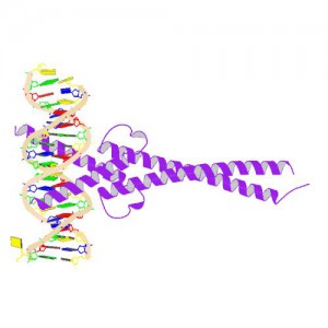
Crystal Structure of MYC MAX Heterodimer bound to DNA ImageSource=RCSB PDB; StructureID=1nkp; DOI=http://dx.doi.org/10.2210/pdb1nkp/pdb;

In 1982, picked up because of its homology to chicken virus genes that could transform cells, MYC became one of the first human genes identified that could drive cellular transformation (1,2). Since that time countless laboratories have prodded and poked the human MYC gene, the MYC protein, their homologs in other animal models, and their transforming viral counterparts.

MYC is a transcription factor and forms heterodimers with a required protein partner, MAX, before binding to the E box sequences of DNA regulatory regions (3). MYC regulates gene expression of many targets through interactions with a host of proteins, often referred to as the MYC Interactome (2).  In fact, MYC is estimated to bind 10–15% of the genome, and it regulates the expression of genes that  are transcribed by by each of the three RNA polymerases (2).

MYC plays a central role in regulating cell growth, proliferation, apoptosis, differentiation and transformation, acting as a central integrator of cellular signals. MYC is tightly regulated at multiple levels from gene expression to protein stability. Dysregulation (usually upregulation) of the amount and stability of Myc protein is observed in many human cancers. Even in cancers in which MYC is not directly involved in transforming cells, its normal expression is often required to support the extracellular matrix and/or vascularization necessary for tumor growth and formation (4).

Because MYC is such a central player cancer pathology, it is an attractive target for cancer therapeutics  (2) .

Continue reading “Targeting MYC: The Need to Study Protein:Protein Interactions in Cells”

Choosing Primary and Control Reporters for Dual-Luciferase Assays

Dual-Reporter Assays give scientists the ability to simultaneously measure two reporter enzymes within a single sample. In dual assays, the activity of an experimental reporter is correlated with the effect of specific experimental conditions, while the activity of a control reporter relays the baseline response, providing an essential internal control that reduces variability caused by differences in cell viability or transfection efficiency. The Nano-Glo® Dual-Luciferase® Reporter (NanoDLR™) Assay provides a choice of two sensitive reporters (firefly and NanoLuc luciferases) for use in dual-assay format. Both reporters give state-of-the-art functionality, raising the question “Which luciferase should be the primary reporter and which should be the control?”

This infographic outlines the various NanoDLR dual-reporter assay choices and the situations where you would choose one format over another. Continue reading “Choosing Primary and Control Reporters for Dual-Luciferase Assays”

Choosing the Best Luciferase Vector for Your Experiment—Now Made Easier with the Vector Selector

4621CAGenetic reporters are used as indicators to study gene expression and cellular events coupled to gene expression. They are widely used in pharmaceutical and biomedical research and also in molecular biology and biochemistry. Typically, a reporter gene is cloned with a DNA sequence of interest into an expression vector that is then transferred into cells. Following transfer, the cells are assayed for the presence of the reporter by directly measuring the reporter protein itself or the enzymatic activity of the reporter protein. A good reporter gene can be identified easily and measured quantitatively when it is expressed (in the organism or cells of interest).

Bioluminescent reporters are ideal for these types of studies because they have a number of important features including:
• Measurements that are almost instantaneous
• Exceptional sensitivity
• A wide dynamic range
• Typically no endogenous activity in host cells to interfere with quantitation

However, one factor that is critical for the success of a bioluminescent reporter assay is the vector.

At Promega we offer several different luciferases as reporters, and the genes for those luciferases are available in a variety of vectors. The vectors may vary in the promoters used or the presence or absence of sequences for rapid degradation. Often seemingly small changes in the vector can make a big difference in the suitability of the vector for a given experimental system. Do you need a reporter with a short half-life to detect rapid changes in gene expression? Are you studying a specifically localized protein? Do you wish to perform a transient or stable transfection?

To make finding the best reporter vector for your experimental system easy, we have developed the Luciferase Reporter Vector Selector. Using this online tool, you can narrow the choices of available vectors by promoter type, application (in vivo imaging, cancer pathway analysis, etc), availability of selectable marker, and type of luciferase.

So, as you design your luciferase reporter experiment, keep in mind this handy tool to help you choose the best luciferase vector for your needs.

Detecting Inhibition of Protein Interactions in vivo

Protein Interactions with NanoBRET

In a paper published in the September 2014 issue of ACS Medicinal Chemistry Letters, researchers from GlaxoSmithKline in the UK and Germany report on the discovery, binding mode and structure:activity relationship of a potent BRPF1 (bromodomain and PHD finger containing protein family) inhibitor. This paper came to our attention as it is one of the first publications to apply Promega NanoBRET technology in an vivo assay that reversibly measures the interaction of protein partners. The technology enabled the identification of a novel inhibitor compound that disrupts the chromatin binding of this relatively unstudied class of bromodomain proteins.

What exactly are bromodomains and why do they matter?
Bromodomains are regions (~100 amino acids) within chromatin regulator proteins that recognize and “read” acetylated lysine residues on histones. These acetylated lysines act as docking stations for regulatory protein complexes via binding of the bromodomain region. Because of their role in chromatin binding and gene regulation, bromodomains have attracted interest as potential targets for anti-cancer treatments. Although some bromodomain-containing proteins (e.g., those in the bromodomain and extraterminal domain (BET) subfamily) are well characterized and have been identified as potential therapeutic targets, others are less well understood.

Continue reading “Detecting Inhibition of Protein Interactions in vivo”

Shedding Light on Protein:Protein Interactions with NanoBRET™ Technique

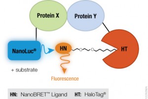
NanoBRET™ TechnologyIf you are trying to investigate protein:protein interactions inside cells, you know how important physiologically relevant results are. If you overload your cells with fusion constructs, your protein interactions may not actually reflect what is going on in the cell, and if your BRET energy donor and acceptor do not have sufficiently separated spectra, you can pick up a fair amount of noise in your experiment. Using the new superbright NanoLuc® Luciferase, and the HaloTag® Technology, we have developed a sensitive BRET system to help you take a better look specific protein interactions that interest you. Promega research scientist, Danette Daniels, describes the system in the Chalk Talk below: