NanoBiT™ Assay: Transformational Technology for Studying Protein Interactions Named a Top 10 Innovation of 2015

21416943-wb-cr-uw-nanobit-pm-page-hero
For three out of the last four years, we have been honored to have one of our key technologies named a Top 10 Innovation by The Scientist. This year the innovative NanoBiT™ Assay (NanoLuc® Binary Technology) received the recognition. NanoBiT™ is a structural complementation reporter based on NanoLuc® Luciferase, a small, bright luciferase derived from the deep sea shrimp Oplophorus gracilirostris.

Using plasmids that encode the NanoBiT complementation reporter, you can make fusion proteins to “report” on protein interactions that you are studying. One of the target proteins is fused to the 18kDa subunit; the other to the 11 amino acid subunit. The NanoBiT™ subunits are stable, exhibiting low self-affinity, but produce an ultra-bright signal upon association. So, if your target proteins interact, the two subunits are brought close enough to each other to associate and produce a luminescent signal. The strong signal and low background associated with a luminescent system, and the small size of the complementation reporter, all help the NanoBiT™ assay overcome the limitations associated with traditional methods for studying protein interactions.

The small size reduces the chances of steric interference with protein interactions. The ultra bright signal, means that even interactions among proteins present in very low amounts can be detected and quantified–without over-expressing large quantities of non-native fusion proteins and potentially disrupting the normal cellular environment. And the NanoBiT™ assay can be performed in real time, in live cells.

The NanoBiT™ assay is already being deployed in laboratories to help advance understanding of fundamental cell biology. You can see how one researcher is already taking full advantage of this innovative technology in the video embedded below:

Visit the Promega web site to see more examples more examples how the NanoBiT™ assay can break through the traditional limitations for studying protein interactions in cells.

You can read the Top 10 article in The Scientist here.
About the Development of an Improved BRET Assay: NanoBRET

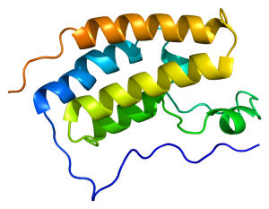
"Protein BRD4 PDB 2oss" by Emw - Own work. Licensed under CC BY-SA 3.0 via Wikimedia Commons - https://commons.wikimedia.org/wiki/File:Protein_BRD4_PDB_2oss.png#/media/File:Protein_BRD4_PDB_2oss.png
“Protein BRD4 PDB 2oss” by Emw – Own work. Licensed under CC BY-SA 3.0 via Wikimedia Commons – https://commons.wikimedia.org/wiki/File:Protein_BRD4_PDB_2oss.png#/media/File:Protein_BRD4_PDB_2oss.png

One of the more exciting reporter molecules technologies available came online in the past year, with the launch of the Promega NanoBRET™ technology. While it’s easy for me, a science writer at Promega, to brag, seriously, this is a very cool protein interactions tool.

A few of the challenges facing protein-protein interactions researchers include:

  • The ability to quantitatively characterize protein-protein interactions
  • Ability to examine protein-protein interactions in situ, in the context of the living cell

A goal of the NanoBRET™ developers was to improve the sensitivity and dynamic range of traditional BRET technology, in order to address these challenges.

In May 2015 these researchers published an article outlining their efforts to create NanoBRET technology in ACS Chemical Biology, in an article entitled, “NanoBRET—A Novel BRET Platform for the Analysis of Protein-Protein Interactions”. Here is a brief look at their work.

Continue reading “About the Development of an Improved BRET Assay: NanoBRET”

Genome Editing and Reporter Technologies Enable Endogenous Pathway High-Throughput Assays

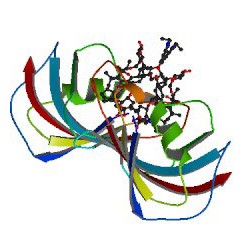
ImageSource=RCSB PDB; StructureID=1qpf; DOI=http://dx.doi.org/10.2210/pdb1qpf/pdb;


This article review was written by guest author, Amy Landreman, in the Cellular Analysis and Proteomics Group at Promega.

Charcot-Marie Tooth (CMT) disease is one of the most common inherited neurological disorders affecting approximately 2.8 million people worldwide. The most common form of CMT, CMT Type 1A, is caused by a 1.5Mb genomic duplication on Chr17 that results in trisomy of the critical myelin gene Peripheral Myelin Protein 22 (Pmp22). The extra copy of Pmp22 results in excessive PMP22 protein causing the neurophathy associated with CMT type 1A. Although there is no way to remove the extra copy of the gene, even subtle decreases in Pmp22 expression have shown promise against this inherited neuropathy in laboratory models.

In a recent paper, Inglese et al. 2014, describe an interesting new approach used to identify compounds that effectively decrease Pmp22 expression using a novel gene editing strategy and reporter-based screen. Their challenge was to create an assay that accurately represented endogenous Pmp22 expression including both transcriptional and post-transcriptional regulatory mechanisms, while maintaining the sensitivity required to detect subtle changes in expression in a loss of signal assay in a format compatible with microtiter 1536-well quantitative high-throughput screening (qHTS). Continue reading “Genome Editing and Reporter Technologies Enable Endogenous Pathway High-Throughput Assays”

For Alphavirus Reporters, Location Matters

Computer-generated model of an alphavirus.

Luminescent reporters offer virologists a convenient way to measure replication of viruses and are also used to image the spread of viruses in vivo in experimental systems. These reporter viruses are useful for evaluating the effects of antiviral drug treatments, testing the efficacy of potential vaccines, and studying the ways in which viruses replicate in the body and cause disease. One challenge in the construction of such reporters is the need to ensure that the reporter molecule itself does not alter the virus in ways that affect its ability to cause disease. Another challenge is maintaining the reporter gene throughout several cycles of viral replication. In smaller viruses, it can be particularly difficult to introduce a reporter gene without compromising the ability of the virus to replicate and cause disease.

A 2014 paper was published in J. Virology comparing the effectiveness of various NanoLuc® luciferase alphavirus reporter constructs. The authors of the study, Chengqun Sun et al. from the University of Pittsburgh, placed these reporter genes in three different locations in the genome of several alphaviruses and compared the effect on their ability to replicate in vitro and in vivo. They also assessed the ability of the luciferase genes to persist during infection of cultured cells and in a mouse model. They showed that the size and location of the reporter had a significant effect on successful replication and persistence. They also showed that the reporters could potentially be integrated at different positions within the genome to study different aspects of viral pathogenesis.

Continue reading “For Alphavirus Reporters, Location Matters”

Using NanoLuc® Luciferase to Study Interactions between Environmental Flavobacteria and Mosquitos

nanoluc

Studies of the larval stages of Aedes triseriatus  (Eastern TreeHole Mosquito) indicate that the “tree hole” habitats in which these larva develop contain diverse microflora including the flavobacteria Elizabethkingia and Chryseobacterium. Extracts from these bacteria have many properties that might affect mosquito health, including antibacterial and anti-fungal activities. Understanding how these bacteria affect larval mosquito development has the potential to inform strategies for mosquito control.

Some initial work has been done by expressing Bacillus larvacidal toxins in some species of Gram-negative bacteria. However, only limited success was achieved using laboratory bacterial strains for such studies. Using environmental flavobacteria might prove to be a more useful approach. However, few molecular tools exist to study environmental flavobacteria. GFP reporters have been used to look at larval feeding, but autofluorescence in the pupae limit the usefulness of GFP-labeled strains for quantitative studies. Furthermore, environmental flavobacteria have unique transcription and translation machinery, and selectable markers and expression plasmids from proteobacteria do not function in these wild strains.

Chen and colleagues set out to generate molecular tools to study Flavobacterium hibernum, a fast-growing bacterium from native mosquito habitats. Their goal was to use these tools to see if A. triseratus larvae ingest and digest these bacteria and to test whether or not F. hibernum can be used to as a vector for larvacidal toxins directed against mosquito larvae. The results of their work were published in Applied and Environmental  Microbiology .

To develop a reporter that avoided issues of autofluorescence background for quantitative studies on the feeding behavior, the researchers turned to NanoLuc® Luciferase, a small, bright luciferase derived from the sea shrimp Oplopphorus gracilirostris. This luciferase has been used in mammalian cells for many kinds of studies, but it has not been used as a reporter in bacterial cells prior to the work of Chen et al. They also looked at work with laboratory flavobacteria strains that used a promoter of outer membrane protein A (PompA) to drive reporter expression as a potential system that might also work with environmental flavobacteria strains.

Continue reading “Using NanoLuc® Luciferase to Study Interactions between Environmental Flavobacteria and Mosquitos”

Another Step Closer to Understanding Epigenetic Gene Regulation

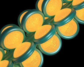
Chromatin fiber

Back when I was a graduate student (more than a few years ago), I remember hearing another student joke that if a member of his thesis committee asked him to explain an unexpected or unusual result, he was going to “blame” epigenetics. At that time, the study of epigenetic gene regulation was in its infancy, and scientists had much to learn about this mysterious regulatory process. Fast forward to today, and you’ll find that scientists know a lot more about basic epigenetic mechanisms, although there is still plenty to learn as scientists discover that the topic is much more complicated than initially thought, as is often the case in science. A recent EMBO Journal article is contributing to our knowledge by shedding light on the role of the TET family of DNA-modifying enzymes in epigenetics (1).

Continue reading “Another Step Closer to Understanding Epigenetic Gene Regulation”