Probing RGS:Gα Protein Interactions with NanoBiT Assays

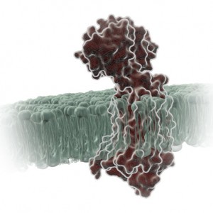
gpcr_in_membrane_on_white2

When I was a post-doc at UT Southwestern, I was fortunate to interact with two Nobel prize winners, Johann Deisenhofer and Fred Gilman.  Johann once helped me move a -80°C freezer into his lab when we lost power in my building. I once replaced my boss at small faculty mixer with a guest speaker and had a drink with Fred Gilman and several other faculty members from around the university. Among the faculty, one professor had a cell phone on his belt, an odd sight in 1995. Fred Gilman asked him what it was and why he had it. It was so his lab could notify him of good results anytime of the day. Fred laughed and told him to get rid of it – if it’s good data, it will survive until morning.

I was reminded of this story when I read a recent paper by Bodle, C.R. et al (1) about the development of a NanoBiT® Complementation Assay (2) to measure interactions of Regulators of G Protein Signaling (RGS) with Gα proteins in cells. (Fred Gilman was the first to isolate a G protein and that led to him being a co-recipient of the Nobel Prize in 1994). The authors created over a dozen NanoBiT Gα:RGS domain pairs and found they could classify different RGS proteins by the speed of the interaction in a cellular context. The interactions were readily reversible with known inhibitors and were suitable for high-throughput screening due to Z’ factors exceeding 0.5. The study paves the way for future work to identify broad spectrum RGS domain:Gα inhibitors and even RGS domain-specific inhibitors. This is the second paper applying NanoBiT Technology to GPCR studies (3).

A Little Background…
A primary function of GPCRs is transmission of extracellular signals across the plasma membrane via coupling with intracellular heterotrimeric G proteins. Upon receptor stimulation, the Gα subunit dissociates from the βγ subunit, initiating the cascade of downstream second messenger pathways that alter transcription (4). The Gα subunits are actually slow GTPases that propagate signals when GTP is bound but shutdown and reassociate with the βγ subunit when GTP is cleaved to GDP. This activation process is known as the GTPase cycle. G proteins are extremely slow GTPases.

Continue reading “Probing RGS:Gα Protein Interactions with NanoBiT Assays”

Shining Light on a Superbug: Clostridium difficile

Antibiotic-resistant bacteria and their potential to cause epidemics with no viable treatment options have been in the news a lot. These “superbugs,” which have acquired genes giving them resistance to common and so-called “last resort” antibiotics, are a huge concern as effective treatment options dwindle. Less attention has been given to an infection that is not just impervious to antibiotics, but is actually enabled by them.

33553646_l

Clostridium difficile Infection (CDI) is one of the most common healthcare-associated infections and a significant global healthcare problem. Clostridium difficile (C. diff), a Gram-positive anaerobic bacterium, is the source of the infection. C. diff spores are very resilient to environmental stressors, such as pH, temperature and even antibiotics, and can be found pretty much everywhere around us, including on most of the food we eat. Ingesting the spores does not usually lead to infection inside the body without also being exposed to antibiotics.

Individuals taking antibiotics are 7-10 times more likely to acquire a CDI. Antibiotics disrupt the normal flora of the intestine, allowing C. diff to compete for resources and flourish. Once exposed to the anaerobic conditions of the human gut, these spores germinate into active cells that embed into the tissue lining the colon. The bacteria are then able to produce the toxins that can cause disease and result in severe damage, or even death.

Continue reading “Shining Light on a Superbug: Clostridium difficile”

Targeting MYC: The Need to Study Protein:Protein Interactions in Cells

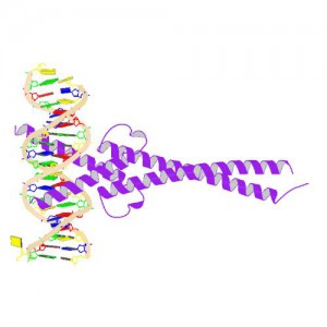
Crystal Structure of MYC MAX Heterodimer bound to DNA ImageSource=RCSB PDB; StructureID=1nkp; DOI=http://dx.doi.org/10.2210/pdb1nkp/pdb;

In 1982, picked up because of its homology to chicken virus genes that could transform cells, MYC became one of the first human genes identified that could drive cellular transformation (1,2). Since that time countless laboratories have prodded and poked the human MYC gene, the MYC protein, their homologs in other animal models, and their transforming viral counterparts.

MYC is a transcription factor and forms heterodimers with a required protein partner, MAX, before binding to the E box sequences of DNA regulatory regions (3). MYC regulates gene expression of many targets through interactions with a host of proteins, often referred to as the MYC Interactome (2).  In fact, MYC is estimated to bind 10–15% of the genome, and it regulates the expression of genes that  are transcribed by by each of the three RNA polymerases (2).

MYC plays a central role in regulating cell growth, proliferation, apoptosis, differentiation and transformation, acting as a central integrator of cellular signals. MYC is tightly regulated at multiple levels from gene expression to protein stability. Dysregulation (usually upregulation) of the amount and stability of Myc protein is observed in many human cancers. Even in cancers in which MYC is not directly involved in transforming cells, its normal expression is often required to support the extracellular matrix and/or vascularization necessary for tumor growth and formation (4).

Because MYC is such a central player cancer pathology, it is an attractive target for cancer therapeutics  (2) .

Continue reading “Targeting MYC: The Need to Study Protein:Protein Interactions in Cells”

Choosing the Best Luciferase Vector for Your Experiment—Now Made Easier with the Vector Selector

4621CAGenetic reporters are used as indicators to study gene expression and cellular events coupled to gene expression. They are widely used in pharmaceutical and biomedical research and also in molecular biology and biochemistry. Typically, a reporter gene is cloned with a DNA sequence of interest into an expression vector that is then transferred into cells. Following transfer, the cells are assayed for the presence of the reporter by directly measuring the reporter protein itself or the enzymatic activity of the reporter protein. A good reporter gene can be identified easily and measured quantitatively when it is expressed (in the organism or cells of interest).

Bioluminescent reporters are ideal for these types of studies because they have a number of important features including:
• Measurements that are almost instantaneous
• Exceptional sensitivity
• A wide dynamic range
• Typically no endogenous activity in host cells to interfere with quantitation

However, one factor that is critical for the success of a bioluminescent reporter assay is the vector.

At Promega we offer several different luciferases as reporters, and the genes for those luciferases are available in a variety of vectors. The vectors may vary in the promoters used or the presence or absence of sequences for rapid degradation. Often seemingly small changes in the vector can make a big difference in the suitability of the vector for a given experimental system. Do you need a reporter with a short half-life to detect rapid changes in gene expression? Are you studying a specifically localized protein? Do you wish to perform a transient or stable transfection?

To make finding the best reporter vector for your experimental system easy, we have developed the Luciferase Reporter Vector Selector. Using this online tool, you can narrow the choices of available vectors by promoter type, application (in vivo imaging, cancer pathway analysis, etc), availability of selectable marker, and type of luciferase.

So, as you design your luciferase reporter experiment, keep in mind this handy tool to help you choose the best luciferase vector for your needs.

Shedding Light on Protein:Protein Interactions with NanoBRET™ Technique

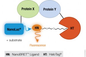
NanoBRET™ TechnologyIf you are trying to investigate protein:protein interactions inside cells, you know how important physiologically relevant results are. If you overload your cells with fusion constructs, your protein interactions may not actually reflect what is going on in the cell, and if your BRET energy donor and acceptor do not have sufficiently separated spectra, you can pick up a fair amount of noise in your experiment. Using the new superbright NanoLuc® Luciferase, and the HaloTag® Technology, we have developed a sensitive BRET system to help you take a better look specific protein interactions that interest you. Promega research scientist, Danette Daniels, describes the system in the Chalk Talk below:

Can Fruit Flies Glow in the Dark?

Fruit fly. Image from morguefile.
Question: How is a fruit fly like a firefly? No, this is not an obvious answer (their names start with the letter “f”) or the beginning of a bad entomology joke. These two organisms may both be winged insects, but as it turns out, what makes the firefly light show such a special treat on summer evenings is something that fruit flies, the bane of the kitchen in the summertime and annoyance for labs near Drosophila researchers, can mimic with a little help from a synthetic luciferin substrate as reported in PNAS. Continue reading “Can Fruit Flies Glow in the Dark?”

Running A Victory Lap For Promega’s Bioluminescence Technologies

Helping scientists design experiments and interpret data is what we do best at Promega Technical Services. This may mean spending time at the bench attempting to reproduce anomalous results or forming a team, perhaps with members of other departments, to brainstorm seemingly intractable experimental road blocks.  Still, for many of us nothing surpasses the experience of meeting these same scientists face to face whether it be on their home turf or at a booth during a tradeshow. PCArticle Continue reading “Running A Victory Lap For Promega’s Bioluminescence Technologies”

Variations on the Two-Hybrid Assay

two-hybrid assays help fit molecules together like puzzle pieces image shows a puzzle

The use of reporter genes for simple analysis of promoter activity (promoter bashing) is a well known practice. However, there are many other elegant applications of reporter technologies. One such application is illustrated in the paper by Zheng et al., published in the Sept. 2008 issue of Cancer Research. These researchers from the Hormel Institute at the University of Minnesota showed that the cyclin-dependent kinase cdk3 phosphorylates the transcription factor ATF1 and enhances its transcriptional and transactivation activity. The observed cdk/ATF1 signaling was shown to have an important role in cell proliferation and transformation. To do this they used several variations of a reporter-based two-hybrid assay.

Continue reading “Variations on the Two-Hybrid Assay”

Why Two Reporters are Better than One

As part of my job I occasionally search the literature for papers citing use of Promega products in new or interesting ways. Any search on dual-luciferase reporters usually generates a lot of returns. A search for dual-luciferase on Highwire press generates over 700 articles from 2009 alone. So why are dual-luciferase reporter assays so widely used? Continue reading “Why Two Reporters are Better than One”