Mass Spec for Glycosylation Analysis of SARS-CoV-2 Proteins Implicated in Host-Cell Entry

The spike protein of the SARS-CoV-2 virus is a very commonly researched target in COVID-19 vaccine and therapeutic studies because it is an integral part of host cell entry through interactions between the S1 subunit of the spike protein with the ACE2 protein on the target cell surface. Viral proteins important in host cell entry are typically highly glycosylated. Looking at the sequence of the SARS-CoV-2 virus, researchers predict that the spike protein is highly glycosylated. In a recent study, researchers conducted a glycosylation analysis of SARS-CoV-2 proteins using mass spec analysis to determine the N-glycosylation profile of the subunits that make up the spike protein.
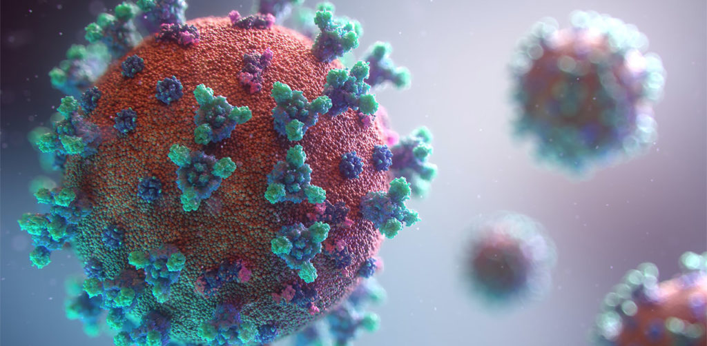
3d model of coronavirus covid-19 showing the spike protein. A recent study performed a glycosylation analysis of SARS-CoV-2 protein.

Glycans assist in protein folding and help the virus avoid immune recognition by the host. Glycosylation can also have an impact on the antigenicity of the virus, as well as potential effects on vaccine safety and efficacy. Mass spectrometry is widely used for viral characterization studies of influenza viruses. Specifically, mass spec has been used to study influenza protein glycosylation, antigen quantification, and determination of vaccine potency.

Continue reading “Mass Spec for Glycosylation Analysis of SARS-CoV-2 Proteins Implicated in Host-Cell Entry”
Understanding the Structure of SARS-CoV-2 Spike Protein

Glycosylation is the process by which a carbohydrate is covalently attached to target macromolecules, typically proteins. This modification serves various functions including guiding protein folding (1,2), promoting protein stability (2), and participating signaling functions (3).

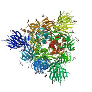
ribbon structure of SARS-CoV-2 protein
Ribbon Structure of SARS-CoV-2 Spike Protein

SARS-CoV-2 utilizes an extensively glycosylated spike (S) protein that protrudes from the viral surface to bind to angiotensin-converting enzyme 2 (ACE2) to mediate host-cell entry. Vaccine development has been focused on this protein, which is the focus of the humoral immune response. Understanding the glycan structure of the SARS-CoV-2 virus spike (S) protein will be critical in the development of glycoprotine-based vaccine candidates.

Continue reading “Understanding the Structure of SARS-CoV-2 Spike Protein”

Glycosyltransferases: What’s New in GT Assays?

In his 2014 blog, “Why We Care About Glycosyltransferases” Michael Curtin, Promega Global Product Manager for Cell Signaling, wrote:

“Glycobiology is the study of carbohydrates and their role in biology. Glycans, defined as ‘compounds consisting of a large number of monosaccharides linked glycosidically’ are present in all living cells; They coat cell membranes and are integral components of cell walls. They play diverse roles, including critical functions in cell signaling, molecular recognition, immunity and inflammation. They are the cell-surface molecules that define the ABO blood groups and must be taken into consideration to ensure successful blood transfusions.

The process by which a sugar moiety is attached to a biological compound is referred to as glycosylation. Protein glycosylation is a form of post-translational modification, which is important for many biological processes and often serves as an analog switch that modulates protein activity. The class of enzymes responsible for transferring the sugar moiety onto proteins is called a glycosyltransferase (GT).”

Continue reading “Glycosyltransferases: What’s New in GT Assays?”

Improved Characterization and Quantification of Complex Cell Surface N-Glycans

MSextractcropped

N-Glycosylation is a common protein post-translational modification occurring on asparagine residues of the consensus sequence asparagine-X-serine/threonine, where X may be any amino acid except proline. Protein N-glycosylation takes place in the endoplasmic reticulum (ER) as well as in the Golgi apparatus.

Approximately half of all proteins typically expressed in a cell undergo this modification, which entails the covalent addition of sugar moieties to specific amino acids. There are many potential functions of glycosylation. For instance, physical properties include: folding, trafficking, packing, stabilization and protease protection. N-glycans present at the cell surface are directly involved in cell−cell or cell−protein interactions that trigger various biological responses.

The standard method used to profile the N-glycosylation pattern of cells is glycoprotein isolation followed by denaturation and/or tryptic digestion of the glycoproteins and an enzymatic release of the N-glycans using PNGase F followed by analysis mass spec. This method has been reported to yield high levels of high-mannose N-glycans that stem from both membrane proteins as well as proteins from the ER.(1,2)

Are you looking for proteases to use in your research?
Explore our portfolio of proteases today.

For those researchers interested in characterizing only cell surface glycans (i.e.,  complex N-glycans)  a recent reference has developed a model system using HEK-292 cells that demonstrates a reproducible, sensitive, and fast method to profile surface N-glycosylation from living cells (3). The method involves standard centrifugation followed by enzymatic release of cell surface N-glycans. When compared to the standard methods the detection and quantification of complex-type N-glycans by increased their relative amount from 14 to 85%.

  1. North, S. J. et al. (2012) Glycomic analysis of human mast cells, eosinophils and basophils. Glycobiology. 2012, 22, 12–22.
  2. Reinke, S. O. et al. (2011) Analysis of cell surface N-glycosylation of the human embryonic
    kidney 293T cell line. J. Carbohydr. Chem.  30, 218–232.
  3. Hamouda, H. et al. (2014) Rapid Analysis of Cell Surface N‑Glycosylation from Living Cells Using Mass Spectrometry. J of Proteome Res. 13, 6144–51.

PNGase F, a Novel Endoglycosidase

11123MA

PNGase F (Cat.# V4831) is a recombinant glycosidase cloned from Elizabethkingia meningoseptica and overexpressed in E. coli, with a molecular weight of 36kD.

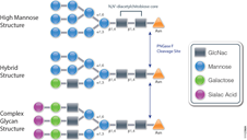
PNGase F catalyzes the cleavage of N-linked oligosaccharides between the innermost GlcNAc and asparagine residues of high mannose, hybrid, and
complex oligosaccharides from N-linked glycoproteins. PNGase F will not remove oligosaccharides containing alpha-(1,3)-linked core fucose,
commonly found on plant glycoproteins.

Applications
Determining whether a protein is in fact glycosylated is the initial step in glycoprotein analysis. Polyacrylamide gel electrophoresis in the
presence of sodium dodecyl sulfate (SDS-PAGE) has become the method of choice as the final step prior to mass spec analysis. Glycosylated proteins often migrate as diffused bands by SDS-PAGE. A marked decrease in band width and change in migration position after treatment with PNGase F is considered evidence of N-linked glycosylation.

Gel based data are often correlated with information obtained from mass spec analysis. Asn-linked type glycans can be cleaved enzymatically by PNGase F yielding intact oligosaccharides and a slightly modified protein in which Asn residues at the site of de-N-glycosylation are converted to Asp, by converting the previously carbohydrate-linked asparagine into an aspartic acid, a monoisotopic mass shift of 0.9840Da is observed. The deglycosylated peptides are then analyzed by tandem mass spectrometry (MS/MS), and software algorithms are used to correlate the experimental fragmentation spectra with theoretical tandem mass spectra generated from peptides in a protein database.

Protease K Protection Assay: Cell Free Expression Application

Microsomal vesicles are used to study cotranslational and initial posttranslational processing of proteins. Processing events such as signal peptide cleavage, membrane insertion, translocation and core glycosylation can be examined by the transcription/translation of the appropriate DNA in the TNT® Lysate Systems when used with microsomal membranes.

The most general assay for translocation makes use of the protection afforded the translocated domain by the lipid bilayer of the microsomal membrane. In this assay protein domains are judged to be translocated if they are observed to be protected from exogenously added protease. To confirm that protection is due to the lipid bilayer addition of 0.1% non-ionic detergent (such as Triton® X-100) solubilizes the membrane and restores susceptibility to the protease.

Are you looking for proteases to use in your research?
Explore our portfolio of proteases today.

Many proteases have proven useful for monitoring translocation in this fashion including Protease K or Trypsin.

The following are examples illustrating this application:

  1. Minn, I. et al. (2009) SUN-1 and ZYG-12, mediators of centrosome-nucleus attachment, are a functional SUN/KASH pair in Caenorhabditis elegans. Mol. Biol. Cell. 20, 4586–95.
  2. Padhan, K. et al. (2007) Severe acute respiratory syndrome coronavirus Orf3a protein interacts with caveolin. J.Gen.Virol. 88, 3067–77.
  3. Tews, B.A. et al. (2007) The pestivirus glycoprotein Erns is anchored in plane in the membrane via an amphipathic helix. J.Biol.Chem. 282, 32730–41.
  4. Pidasheva, S. et al. (2005) Impaired cotranslational processing of the calcium-sensing receptor due to signal peptide missense mutations in familial hypocalciuric hypercalcemia. Hum. Mol. Gen. 14, 1679–90.
  5. Smith, D. et al. (2002) Exogenous peptides delivered by ricin require processing by signal peptidase for transporter associated with antigen processing-independent MHC class I-restricted presentation. J. Immun. 169, 99–107.