Ancient Retroviruses and Modern Cancer: Role of Endogenous Retroviruses in Transcriptional Changes in Tumor Cells 

Approximately 30 million years ago, a retrovirus integrated into the germline of a common ancestor of baboons, gorillas, chimpanzees and humans. That endogenous retrovirus, now known as gammaretrovirus human endogenous retrovirus 1 (HERV-1), may provide clues about the aberrant regulation of gene transcription that enables tumor cells to grow and survive.  

Understanding the Mechanism Behind Cancer Gene Expression 

Scientists have long described the striking differences in gene expression, signaling activity and metabolism between cancer cells and normal cells, but the underlying mechanisms that cause these differences are not fully understood. In a recent Science Advances article, published by Ivancevic et al., researchers from the University of Colorado, Boulder; the University of Colorado Anschutz Medical Campus, and the University of Colorado School of Medicine report their efforts to identify endogenous retrovirus elements that might be part of the answer to the complex question of what biological events are responsible for the changes in gene expression in cancer cells.  

The researchers hypothesized that transposable elements (TEs), specifically those associated with endogenous retroviruses could be involved in cancer-specific gene regulation.  Endogenous retroviruses (ERVs) are the remnants of ancient retroviral infections that have integrated into the germline of the host. 

The transposable element LTR10, derived from an endogenous retrovirus, can alter gene expression in a number of cancers. Artist's conception of an invasive cancer cell.

Identifying Endogenous Retrovirus Elements That Affect Cancer Gene Expression 

Continue reading “Ancient Retroviruses and Modern Cancer: Role of Endogenous Retroviruses in Transcriptional Changes in Tumor Cells “

Insects and Science: Optimizing Work with Sf9 Insect Cells

Insects are a keystone species in the animal kingdom, often providing invaluable benefits to terrestrial ecosystems and useful services to mankind. While many of them are seen as pests (think mosquitos), others are important for pollination, waste management, and even scientific research.

Insect biotechnology, or the use of insect-derived molecules and cells to develop products, is applied in a diverse set of scientific fields including agricultural, industrial, and medical biotechnology. Insect cells have been central to many scientific advances, being utilized in recombinant protein, baculovirus, and vaccine and viral pesticide production, among other applications (5).

Therefore, as the use of insect cells becomes more widespread, understanding how they are produced, their research applications, and the scientific products that can be used with them is crucial to fostering further scientific advancements.

Primary Cell Cultures and Cell Lines

Cell culture - Cell lines - Insect Cells

In general, experimentation with individual cells, rather than full animal models, is advantageous due to improved reproducibility, decreased space requirements, less ethical concerns, and a reduction in expense. This makes primary cell cultures and cell lines essential contributors to basic scientific research.

Continue reading “Insects and Science: Optimizing Work with Sf9 Insect Cells”

Cellular Senescence and Cancer Therapy: Overcoming Immortality?

At the time of writing this post, no scientist had yet discovered the secret to immortality. In our world, we’ve come to accept that living things are born, grow old and die—the circle of life.

And yet, for many years, life scientists believed that the circle of life did not apply to our constituent cells when cultured in a laboratory. That is, cultured normal human cells were immortal, and they would continue to grow and proliferate forever, as long as they were provided with the necessary nutrients.

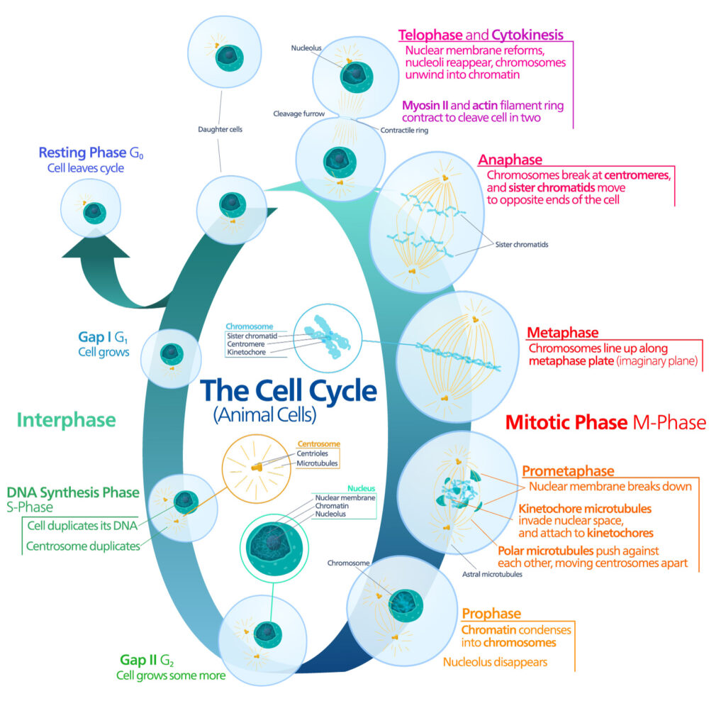
The animal cell cycle. Image by Kelvinsong; made available under the Creative Commons CC0 1.0 Universal Public Domain Dedication

Pioneering work published in 1961 by Leonard Hayflick and Paul Moorhead challenged that theory (reviewed in 1). Their research showed that normal cells in culture have a finite capacity to replicate. After they reach a certain number of replicative cycles, cells stop dividing. Hayflick and Moorhead made the important distinction between normal human cells and cultured cancer cells, which are truly immortal. In later years, the limit to the number of replicative cycles normal human cells can undergo became known as the Hayflick limit. Although some scientists still express skepticism about these findings, the Hayflick limit is widely recognized as a fundamental principle of cell biology.

Continue reading “Cellular Senescence and Cancer Therapy: Overcoming Immortality?”

Out-FOXOing High-Stage Neuroblastoma

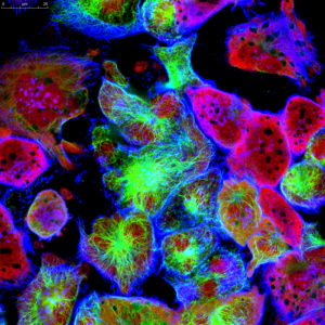
Fluorescence microscopy of neuroblastoma cells.

In recent years, scientists have been hot on the trail of transcription factor FOXO3, tracing its involvement in various tumor-centric activities comprising the many trademarks of cancer, from drug resistance to metastasis to tumor angiogenesis.

FOXO3 is a member of the O sub-class of the forkhead box family of transcription factors. The forkhead box (FOX) family is characterized by a fork head DNA-binding domain (DBD), comprised of around 100 amino acids. They have also proven themselves to be a family of many hats, functioning in diverse roles ranging from metabolism, immunology, cell-cycle control, development, as well as cancer (1). The forkhead box O (FOXO) sub-class alone has demonstrated involvement in a variety of cellular outcomes, from drug resistance and longevity to apoptosis induction.

Due to its pro-apoptotic and anti-proliferative proclivity, FOXO3 has been previously identified as a tumor suppressor gene. However, more and more studies have begun to flip the narrative on FOXO3, portraying it more as a devoted henchman, due to its roles in drug and radiotherapy resistance, cell-cycle arrest and long-term maintenance of leukemia-initiating stem cells in a variety of cancer types, including breast cancer, pancreatic cancer, glioblastoma, and both acute and chronic myeloid leukemia.

Continue reading “Out-FOXOing High-Stage Neuroblastoma”

How to Reduce Cell Culture Variability

Scenario 1: Jake needs a flask of MCF-7 cells for an assay, so he sends an email to the graduate student listserv asking for cells. Melissa replies that she has an extra flask of cells that she could share. Jake happily accepts the cells and begins his experiment.

Scenario 2: Michael passaged his cells yesterday and, according to the protocol, was supposed to plate cells today for treatment. However, his previous experiments were delayed, so he decides to plate them tomorrow instead. The cells look healthy, so it should be ok.

What is wrong with the above scenarios? These actions may seem harmless, but they could be the cause of variability, leading to irreproducible results.

Continue reading “How to Reduce Cell Culture Variability”

A Better Way to Understand How and Why Cells Die

Real-time, up-to-the-minute access to information provides new opportunities for scientists to monitor cellular events in ever more meaningful ways. Real-time cytotoxicity and cell viability assay reagents now allow constant monitoring of cell health status without the need to lyse or remove aliquots from plates for measurement. With a real-time approach, data can be collected from cell cultures or microtissues at multiple time points after addition of a drug compound or other event, and the response to treatment continually observed.

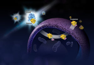
The CellTox™ Green assay is a real-time assay that monitors cytotoxicity using a fluorescent DNA binding dye, which binds DNA released from cells upon loss of membrane integrity. The dye cannot enter intact, live cells and so fluorescence only occurs upon cell death, correlating with cytotoxicity. Here’s a quick overview showing how the assay works:

More Data Using Fewer Samples and Reagents
The ability to continually monitor cytotoxicity in this way makes it easy to conduct more than one type of analysis on a single sample. Assays can be combined to determine not only the timing of cytotoxicity, but to also understand related events happening in the same cell population. As long as the readouts can be distinguished from one another multiple assays can be performed in the same well, providing more informative data while using less cells, plates and reagents.

Combining assays in this way can reveal critical information regarding mechanism of cell death. For example, assay combinations can be used to determine whether cells are dying from apoptosis or necrosis, or to distinguish nonproliferation from cell death. Combining CellTox Green with an endpoint luminescent caspase assay or a real-time apoptosis assay allows you to determine whether observed cytotoxic effects are due to apoptosis. Cytotoxic and anti-proliferative effects can be distinguished by combining the cytotoxicity assay with a luminescent or fluorescent cell viability assay.

Continue reading “A Better Way to Understand How and Why Cells Die”

Reveal More Biology: How Real-Time Kinetic Cell Health Assays Prove Their Worth

What if you could uncover a small but significant cellular response as your population of cells move toward apoptosis or necrosis? What if you could view the full picture of cellular changes rather than a single snapshot at one point? You can! There are real-time assays that can look at the kinetics of changes in cell viability, apoptosis, necrosis and cytotoxicity—all in a plate-based format. Seeking more information? Multiplex a real-time assay with endpoint analysis. From molecular profiling to complementary assays (e.g., an endpoint cell viability assay paired with a real-time apoptosis assay), you can discover more information hidden in the same cells during the same experiment.
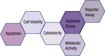
Whether your research involves screening a panel of compounds or perturbing a regulatory pathway, a more complete picture of cellular changes gives you the benefit of more data points for better decision making. Rather than assessing the results of your experiment using a single time point, such as 48 hours, you could monitor cellular changes at regular intervals. For instance, a nonlytic live-cell reagent can be added to cultured cells and measurements taken repeatedly over time. Pairing a real-time cell health reagent with a detection instrument that can maintain the cells at the correct temperature means you can automate the measurements. These repeated measurements over time reveal the kinetic changes in the cells you are testing, giving a real-time status update of the cellular changes from the beginning to the end of your experiment. Continue reading “Reveal More Biology: How Real-Time Kinetic Cell Health Assays Prove Their Worth”

Piecing the Puzzle Together: Using Multiple Assays to Better Understand What Is Happening with Your Cells

You often need several pieces of information to really understand what is happening within a cell or population of cells. If your cells are not proliferating, are they dying? Or, are you seeing cytostasis? If they are dying, what is the mechanism? Is it apoptosis or necrosis? If you are seeing apoptosis, what is the pathway: intrinsic or extrinsic?

If you are measuring expression of a reporter gene and you see a decrease in expression, is that decrease due to transfection inefficiencies, cytotoxicity, or true down regulation of your reporter gene?

To investigate these multiple parameters, you can run assays in parallel, but that requires more sample, and sample isn’t always abundant.

Multiplexing assays allows you to obtain information about multiple parameters or events (e.g., reporter gene expression and cell viability; caspase-3 activity and cell viability) from a single sample. Multiplexing saves sample, saves time and gives you a more complete picture of the biology that is happening with your experimental sample.

What information do you need about your cells to complete the picture?

Multiplexing assay reagents to measure biomarkers in the same sample has often been considered an application only accomplished with antibodies or dyes and sophisticated detection instrumentation. However, Promega has developed microwell plate based assays for cells in culture that allow multiplexed detection of biomarkers in the same sample well using standard multimode multiwell plate readers. Continue reading “Piecing the Puzzle Together: Using Multiple Assays to Better Understand What Is Happening with Your Cells”

Black Raspberry Extract May Lead to Tomorrow’s Cancer Preventative

Black raspberry shrubs. Copyright Sara KlinkWhen deciding which varieties of fruit to cultivate, I chose to plant black raspberries on my small suburban lot. They grow wild in Wisconsin, but fighting through swarms of mosquitos, brush and thorns to pick berries was not my idea of fun. For the last two years, I have received a large crop of juicy black berries that I enjoy eating fresh or process into black raspberry jam to spread on toast. Therefore, I was interested to learn that black raspberries have demonstrated cancer preventative properties in animal models of chemically induced oral and colon cancers as well as cultured oral cancer cells. Due to similarities between oral and cervical cancers, researchers recently tested if the beneficial effects of this berry could extend to human cervical cancer cells. Continue reading “Black Raspberry Extract May Lead to Tomorrow’s Cancer Preventative”

Screening for Drug-Drug Interactions with PXR and CYP450 3A4 Activation

The pregnane X receptor (PXR) is a nuclear receptor known to regulate expression of cytochrome P450 (CYP450) drug-metabolizing enzymes (1). PXR has even been designated the “master xenosensor” due to its ability to upregulate cellular levels of a variety of drug-metabolizing enzymes in response to drugs and foreign chemicals. Elevated levels of CYP450 enzymes can elicit alterations in the pharmacokinetics of co-administered drugs, which can result in adverse drug-drug interactions (DDI) or diminished bioavailability. By assessing PXR activation and CYP450 enzyme induction early in the drug development process, many companies hope to reduce late-stage clinical failures and minimize the high costs associated with bringing a new drug to market.

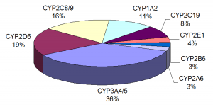
Proportion of drugs metabolized by different CYPs

A paper by Shukla et al. (2) examined over 2,800 clinically used drugs for their ability to activate human PXR (hPXR) and rat PXR (rPXR), induce human cytochrome P450 3A4 enzyme (CYP3A4) at the cellular level, and bind hPXR at the protein level. Several studies have identified PXR as playing a key role in regulating the expression of CYP3A4, an enzyme involved in the metabolism of more than 50% of all drugs prescribed in humans. Since PXR activation and CYP3A4 induction have an impact on drug metabolism and pharmacokinetics, the authors wanted to obtain data that would be valuable in understanding structure-activity relationships (SARs), the connection between chemical structure and biological activity, when prioritizing new molecular entities (NMEs) for further in vitro and in vivo studies.

Continue reading “Screening for Drug-Drug Interactions with PXR and CYP450 3A4 Activation”