Glo-ing Above and Beyond: Simplifying Science with MyGlo Reagent Reader

Introduction

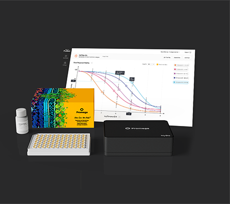
When it comes to laboratory tools, few things resonate more than the experiences of researchers who rely on them daily. At the University of Cincinnati the MyGlo Reagent Reader has quickly become an indispensable lab companion, due to its compact design, affordability, and intuitive interface with tailored apps for Promega assays. But what truly sets the MyGlo Reagent Reader apart is how it empowers scientists to focus on their research.

Take Ipsita Kundu, a third-year PhD student at the University of Cincinnati working in Dr. Tim Phoenix’s lab. The Phoenix lab, dedicated to studying innovative brain tumor therapies, faced challenges with their outdated luminescence reader. They needed an affordable, reliable solution to streamline Ipsita’s experiments without compromising accuracy or efficiency.

The MyGlo Reagent Reader was the answer. This blog highlights how this integrated solution is redefining laboratory workflows, enabling researchers to maximize productivity and maintain focus on groundbreaking discoveries. Let’s delve into Ipsita’s story and explore how MyGlo transformed her research.

Continue reading “Glo-ing Above and Beyond: Simplifying Science with MyGlo Reagent Reader”

The Greatness of Glycogen: A Central Storage Molecule in Energy Metabolism

Introduction

Glycogen is a fundamental molecule in energy metabolism, serving as the critical storage form of glucose that supports cellular health and energy homeostasis. As a polysaccharide, glycogen is essential for maintaining stable energy levels, particularly during periods of fasting and physical exertion. This article will examine glycogen’s synthesis, storage, and utilization, along with its broader significance in human health and disease. Understanding glycogen’s role can provide valuable insight into energy regulation and metabolic health.

Continue reading “The Greatness of Glycogen: A Central Storage Molecule in Energy Metabolism”

The Benefits of BCAAs: Branched-Chain Amino Acids in Health and Disease

Introduction

Branched-chain amino acids (BCAAs) are essential nutrients that play a significant role in muscle metabolism and overall health. Comprised of leucine, isoleucine, and valine, BCAAs cannot be synthesized by the body and must be obtained through diet. Recent research has highlighted how the metabolic pathways are influenced by BCAAs, such as their ability to activate mTOR signaling, which is vital for muscle protein synthesis (Choi, 2024). Beyond muscle growth, BCAAs may support cognitive function and metabolic health, with ongoing research exploring their broader benefits in disease management. This article explores the diverse roles of BCAAs and their impact on health and diseases

Continue reading “The Benefits of BCAAs: Branched-Chain Amino Acids in Health and Disease”

The Brilliance of BHB: A Key Ketone Body in Metabolic Health

Introduction

β-Hydroxybutyrate (BHB), the most abundant ketone body, is a crucial molecule that sustains energy production during periods of glucose deprivation. Whether you are fasting, adhering to a ketogenic diet, or simply interested in metabolic flexibility, BHB offers key insights into how our bodies adapt to alternative energy sources. This article will delve into how BHB is produced, the diverse roles it plays, and its implications for health and disease.

Continue reading “The Brilliance of BHB: A Key Ketone Body in Metabolic Health”

Decoding the NAD+/NADH Ratio and Its Crucial Role in Cell Health

Nicotinamide adenine dinucleotide (NAD) exists in two forms in the cell: NAD+ (oxidized) and NADH (reduced). This molecule plays a pivotal role in metabolic processes, serving as a key electron carrier in the redox reactions that drive cellular metabolism. The balance between these two forms, commonly expressed as the NAD+/NADH ratio, is crucial for maintaining cellular function and the intracellular redox state. This article explores the significance of this ratio, how it impacts cellular processes, and the consequences of NAD+/NADH ratio dysregulation.

Continue reading “Decoding the NAD+/NADH Ratio and Its Crucial Role in Cell Health”

Cellular Senescence and Cancer Therapy: Overcoming Immortality?

At the time of writing this post, no scientist had yet discovered the secret to immortality. In our world, we’ve come to accept that living things are born, grow old and die—the circle of life.

And yet, for many years, life scientists believed that the circle of life did not apply to our constituent cells when cultured in a laboratory. That is, cultured normal human cells were immortal, and they would continue to grow and proliferate forever, as long as they were provided with the necessary nutrients.

The animal cell cycle. Image by Kelvinsong; made available under the Creative Commons CC0 1.0 Universal Public Domain Dedication

Pioneering work published in 1961 by Leonard Hayflick and Paul Moorhead challenged that theory (reviewed in 1). Their research showed that normal cells in culture have a finite capacity to replicate. After they reach a certain number of replicative cycles, cells stop dividing. Hayflick and Moorhead made the important distinction between normal human cells and cultured cancer cells, which are truly immortal. In later years, the limit to the number of replicative cycles normal human cells can undergo became known as the Hayflick limit. Although some scientists still express skepticism about these findings, the Hayflick limit is widely recognized as a fundamental principle of cell biology.

Continue reading “Cellular Senescence and Cancer Therapy: Overcoming Immortality?”

Save Precious Time with Same-Well Multiplexing

Scientist performing a multi-well assay. Same-well multiplexing enables you to look at one event from several perspectives.

A graduate student believes he has mastered the art of “the assay”. No need to run duplicates, he knows exactly which one will get him the answers he needs right away.  

To challenge this, his PI proposes an exercise. He asks of the graduate student, “What happens when you treat cells with doxorubicin?”

The graduate student raises his cells, treats them accordingly, and decides to run a cell viability assay to determine their fate. He returns to the PI with the final verdict: his cells are dead.

The PI takes a look at the data and asks the graduate student to repeat the experiment with an additional assay for cytotoxicity―but the cytotoxicity assay shows that the cell membranes are intact, which only puzzles the graduate student. The PI asks him to run a third assay for apoptosis, and when the student does so, it becomes clear that the cells are dying.

The PI uses this opportunity to make his point: “Now do you see why I ask for more than one assay?”

Continue reading “Save Precious Time with Same-Well Multiplexing”

NAD: A Renaissance Molecule and its Role in Cell Health

NAD is a pyridine nucleotide. It provides the oxidation and reduction power for generation of ATP by mitochondria. For many years it was believed that the primary function of NAD/NADH in cells was to harness and transfer energy  from glucose, fatty and amino acids through pathways like glycolysis, beta-oxidation and the citric acid cycle.

Promega NAD/NADH-Glo system and how to prepare samples for  identification of NAD or NADH.
Promega NAD/NADH-Glo system and how to prepare samples for identification of NAD or NADH.

However NAD also is recognized as an important cell signaling molecule and substrate. The many regulatory pathways now known to use NAD+ in signaling include multiple aspects of cellular homeostasis, energy metabolism, lifespan regulation, apoptosis, DNA repair and telomere maintenance.

This resurrection of NAD importance is due in no small part to the discovery of NAD-using enzymes, especially the sirtuins.

Continue reading “NAD: A Renaissance Molecule and its Role in Cell Health”

A Tale of Two Toxins: the mechanisms of cell death in Clostridium difficile infections

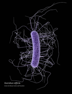
When someone is admitted to a hospital for an illness, the hope is that medical care and treatment will help them them feel better. However, nosocomial infections—infections acquired in a health-care setting—are becoming more prevalent and are associated with an increased mortality rate worldwide. This is largely due to the misuse of antibiotics, allowing some bacteria to become resistant. Furthermore, when an antibiotic wipes out the “good” bacteria that comprise the human microbiome, it leaves a patient vulnerable to opportunistic infections that take advantage of disruptions to the gut microbiota.

One such bacteria, Clostridium difficile, is of growing concern world-wide since it is resistant to many different antibiotics. When a patient is treated with an antibiotic, C. difficile can thrive in the intestinal tract without other bacteria populating the gut. C. difficile infection is the leading cause of antibiotic-associated diarrhea. While symptoms can be mild, aggressive infection can lead to pseudomembranous colitis—a severe inflammation of the colon which can be life-threatening.

C. difficile causes disease by releasing two large toxins, TcdA and TcdB. Understanding the role these toxins play in colonic disease is important for treatment strategies. However, most published research data only report the effects of the toxins independently. A 2016 study demonstrated a method of comparing the toxins side-by-side using the same time points and cell assays to investigate the role each toxin plays in the cell death that leads to disease of the colon. Continue reading “A Tale of Two Toxins: the mechanisms of cell death in Clostridium difficile infections”

A Better Way to Understand How and Why Cells Die

Real-time, up-to-the-minute access to information provides new opportunities for scientists to monitor cellular events in ever more meaningful ways. Real-time cytotoxicity and cell viability assay reagents now allow constant monitoring of cell health status without the need to lyse or remove aliquots from plates for measurement. With a real-time approach, data can be collected from cell cultures or microtissues at multiple time points after addition of a drug compound or other event, and the response to treatment continually observed.

The CellTox™ Green assay is a real-time assay that monitors cytotoxicity using a fluorescent DNA binding dye, which binds DNA released from cells upon loss of membrane integrity. The dye cannot enter intact, live cells and so fluorescence only occurs upon cell death, correlating with cytotoxicity. Here’s a quick overview showing how the assay works:

More Data Using Fewer Samples and Reagents
The ability to continually monitor cytotoxicity in this way makes it easy to conduct more than one type of analysis on a single sample. Assays can be combined to determine not only the timing of cytotoxicity, but to also understand related events happening in the same cell population. As long as the readouts can be distinguished from one another multiple assays can be performed in the same well, providing more informative data while using less cells, plates and reagents.

Combining assays in this way can reveal critical information regarding mechanism of cell death. For example, assay combinations can be used to determine whether cells are dying from apoptosis or necrosis, or to distinguish nonproliferation from cell death. Combining CellTox Green with an endpoint luminescent caspase assay or a real-time apoptosis assay allows you to determine whether observed cytotoxic effects are due to apoptosis. Cytotoxic and anti-proliferative effects can be distinguished by combining the cytotoxicity assay with a luminescent or fluorescent cell viability assay.

Continue reading “A Better Way to Understand How and Why Cells Die”