Looking Back: Cell-Free Expression Systems Helped to Characterize Proteins Involved in Hypoxia Response

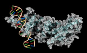
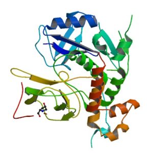
Structur of a HIF-1a-pVHL-ElonginB-ElonginC complex
Structure of a HIF-1a-pVHL-ElonginB-ElonginC complex

William G. Kaelin Jr., Sir Peter J. Ratcliffe and Gregg L. Semenza were awarded the 2019 Nobel Prize in Physiology or Medicine for their discoveries of how cells sense and adapt to oxygen availability.

Kaelin and Ratcliffe’s labs focused their efforts on the transcription factor HIF (hypoxia-inducible factor). This transcription factor is critical in the cellular adaptation of to changes in oxygen availability.

When oxygen levels are elevated cells contain very little HIF. Ubiquitin is added to the HIF protein via the VHL complex and it is degraded in the proteasome.  When oxygen levels are low (hypoxia) the amount of HIF increases.

In 2001 both groups published articles characterizing the interaction between VHL and HIF, and these articles were referenced by the Nobel Prize Organization in their press release about this year’s award. (1,2). Both studies demonstrated that under the normal oxygen conditions hydroxylation of proline residue P564 enabled VHL to recognize and bind to HIF.

The use of cell free expression (i.e., TNT Coupled Transcription/Translation System) by both labs was key in the characterization of the VHL:HIF interaction The labs utilized HIF and VHL 35-S labeled proteins generated via the TNT system under both normal or in a hypoxic work station to:

  • Determine the affect of ferrous chloride and cobaltous chloride on the interaction
  • Map the specific region of HIF required for the interaction to occur (556-574)
  • Determine the effect of HIF point mutations on the interaction
  • Use synthetic peptides to block the interaction
  • Conclude that a factor in mammalian cells was necessary for the interaction to occur.

Literature Cited

  1. Ivan, M et al. (2001) HIF Targeted for VHL-Mediated Destruction by Proline Hydroxylation: Implications for O2 Sensing. Science 292: 464–67.
  2. Jaakkola, P. et al. (2001) Targeting of HIF-α to the von Hippel-Lindau Ubiquitylation of Complex by O2– Regulated Prolyl Hydroxylation. Science 202, 468–72 .
Related Posts

Characterizing Compound Binding in Cell-Free Systems

Dioxins (e.g., 2,3,7,8-Tetrachlorodibenzo-p-dioxin, TCDD) and related compounds (DRCs) are persistent environmental pollutants that gradually accumulate through the food chain, mainly in the fatty tissues of animals. Dioxins are highly toxic and can cause reproductive and developmental problems, damage the immune system, interfere with hormones and also cause cancer. This broad range of toxic and biological effects of DRCs is mostly mediated by the aryl hydrocarbon receptor (AHR).

In animal cells, DRCs bind to AHR in the cytoplasm and then translocate into the nucleus, where they affect the transcription of multiple target genes, including xenobiotic-metabolizing enzymes, such as CYP1A isozymes. AHR is also involved in immune system maintenance, protein degradation and cell proliferation.

The jungle crow (Corvus macrorhynchos) has been considered a suitable indicator for monitoring environmental chemicals such as DRCs. While mammals only have one AHR form, avian species have multiple AHR isoforms such as AHR1 and AHR2. To unveil the functional diversity of multiple avian AHR isoforms in terms of their contribution to responses to DRCs a recent study by Kim et al. investigated the molecular and functional characteristics of jungle crow AHR isoforms, cAHR1 and jcAHR2 (1).

cAHR1 and jcAHR2 proteins were synthesized using AHR proteins were synthesized using the TnT Quick-Coupled Reticulocyte Lysate System  to examine whether these jcAHRs have the potential to bind to TCDD. TCDD-binding affinity of the in vitro-expressed jcAHR protein was analyzed using the velocity sedimentation assay with a sucrose gradient.

The results demonstrate that both jcAHR1and jcAHR2 are capable of binding to TCDD.

Reference
Kim, E-Y (2019) The aryl hydrocarbon receptor 2 potentially mediates cytochrome P450 1A induction in the jungle crow (Corvus macrorhynchos). Ecotoxicology and Environmental Safety 171. 99–111

Cell Free Application: Characterization of Long Non-coding RNA Inhibition of Transcription

Long noncoding RNAs have been shown to regulate chromatin states, transcriptional activity and post transcriptional activity (1). Only a few studies have observed long non-coding RNAs modulating the translational process (2). The noncoding RNA BC200 has been shown to inhibit translation by interacting with the translation initiation factors, eIF4A and eIF4B.

To characterize how BC200 translational inhibition could be controlled,  a variety of RNAs were transcribed/translated in vitro using the TNT system (Cat. #L4610) from Promega. To each transcription/translation reaction, BC900 RNA, hnRNPE1 and hnRNE2 proteins were added. Inhibition of BC200 activity was noted when proteins were successful expressed (3).

Literature Cited

  1.  Sosinska, P et.al. (2015) Intraperitoneal invasiveness of ovarian cancer from the cellular and molecular perspective. Ginekol. Pol. 86, 782–86.
  2. Geisler, S. and Coller, J. (2013) RNA in unexpected places: long non-coding RNA functions in diverse cellular contexts. Nat.Rev. Mol. Cell. Bio. 14,699–12.
  3. Jang, S. et. al. (2017) Regulation of BC200 RNA-mediated translation inhibition by hnRNP E1 and E2. FEBS Letters. 591, 393–5.

Luciferase Immunoprecipitation System Assay (LIPS): Expression of Luciferase Antigen using TNT Transcription/Translation Kit

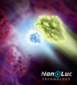
NanoLuc dual reporters
Illustration showing NanoLuc and firefly luciferase reporters.

The luciferase immunoprecipitation system (LIPS) assay is a liquid phase immunoassay allowing high-throughput serological screening of antigen-specific antibodies. The immunoassay involves quantitating serum antibodies by measuring luminescence emitted by the reporter enzyme Renilla luciferase (Rluc) fused to an antigen of interest. The Rluc-antigen fusion protein is recognized by antigen-specific antibodies, and antigen-antibody complexes are captured by protein A/G beads that recognize the Fc region of the IgG antibody (1).

In a recent publication (2), this assay was used to assess the presence of autoantibodies against ATP4A and ATP4B subunits of parietal cells H+, K+-ATPase in patients with atrophic body gastritis and in controls. Continue reading “Luciferase Immunoprecipitation System Assay (LIPS): Expression of Luciferase Antigen using TNT Transcription/Translation Kit”

Cell Free Expression Application: In vitro degradation assay

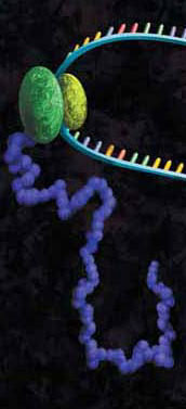
A protein chain being produced from a ribosome.

Researchers and clinicians are fairly certain that all cervical cancers are caused by Human Papillomavirus (HPV) infections, and that HPV16 and HPV18 are responsible for about 70% of all cases. HPV16 and HPV18 have also been shown to cause almost half the vaginal, vulvar, and penile cancers, while about 85% of anal cancers are also caused by HPV16.

E6 is a potent oncogene of HR-HPVs, and its role in progression to malignancy continues to be explored. The E6 oncoprotein of HPV can promote viral DNA replication through several pathways. It forms a complex with human E3-ubiquitin ligase E6-associated protein (E6AP), which can in turn target the p53 tumor-suppressor protein, leading to its ubiquitin-mediated degradation. In particular, E6 from HR-HPVs can block apoptosis, activate telomerase, disrupt cell adhesion, polarity and epithelial differentiation, alter transcription and G-protein signaling, and reduce immune recognition of HPV-infected cells.

In a recent publication a new procedure generated a stable, unmutated HPV16 E6 protein (1). Continue reading “Cell Free Expression Application: In vitro degradation assay”

Protein:DNA Interactions—High-Throughput Analysis

Protein-DNA interactions are fundamental processes in gene regulation in a living cells. These interactions affect a wide variety of cellular processes including DNA replication, repair, and recombination. In vivo methods such as chromatin immunoprecipitation (1) and in vitro electrophoretic mobility shift assays (2) have been used for several years in the characterization of protein-DNA interactions. However, these methods lack the throughput required for answering genome-wide questions and do not measure absolute binding affinities. To address these issues a recent publication (3) presented a high-throughput micro fluidic platform for Quantitative Protein Interaction with DNA (QPID). QPID is an microfluidic-based assay that cam perform up to 4096 parallel measurements on a single device.

The basic elements of each experiment includes oligonucleotides that were synthesized and hybridized to a Cy5-labeled primer and extended using Klenow. All transcription factors that were evaluated contained a 3’HIS and 5’ cMyc tag and were expressed in rabbit reticulocyte coupled transcription and translation reaction (TNT® Coupled Reticulocyte Lysate). Expressed proteins are loaded onto to the QIPD device and immobilized. In the DNA binding assay the fluorescent DNA oligonucleotides are incubated with the immobilized transcription factors and fluorescent images taken. To validate this concept the binding of four different transcription factor complexes to 32 oligonucleotides at 32 different concentrations was characterized in a single experiment. In a second application, the binding of ATF1 and ATF3 to 128 different DNA sequences at different concentrations were analyzed on a single device.

Literature Cited

  1. Ren, B. et al. (2007) Genome-wide mapping of in vivo protein-DNA binding proteins. Science 316, 1497–502.
  2. Garner, M.M. (1981) A gel electrophoresis method for quantifying the binding of proteins to specific DNA regions. Nuc. Acids. Res. 9, 3047-60.
  3. Glick,Y et al. (2016) Integrated microfluidic approach for quantitative high throughput measurements of transcription factor binding affinities. Nuc. Acid Res. 44, e51.

Cell-free Expression: A System for Every Need

6634MA

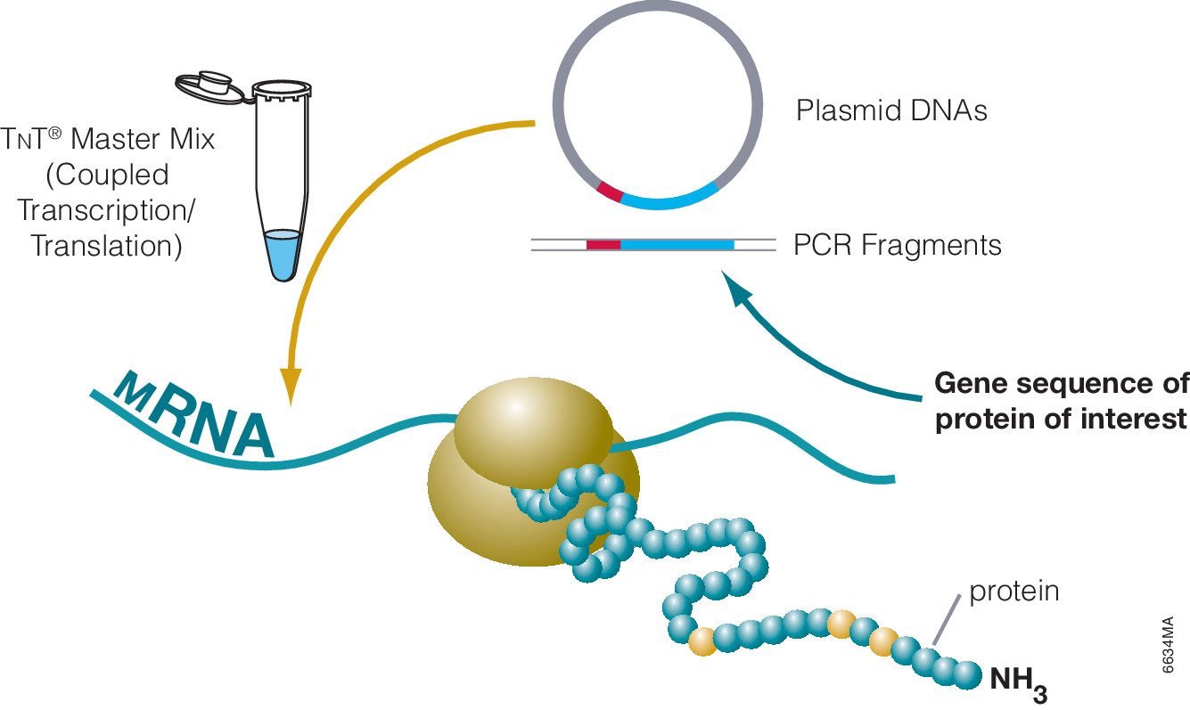
Cell-free protein expression is a simplified and accelerated avenue for the transcription and/or translation of a specific protein in a quasi cell environment. An alternative to slower, more cumbersome cell-based methods, cell-free protein expression methods are simple and fast and can overcome toxicity and solubility issues sometimes experienced in traditional E. coli expression systems. Continue reading “Cell-free Expression: A System for Every Need”

Compound Screening Using Cell-Free Protein Expression Systems

A protein chain being produced from a ribosome.
Both prokaryotic and eukaryotic cell-free protein expression systems have found great utility in efforts to screen organic compounds for inhibition of the basic cellular functions of transcription and translation, common targets for antibiotic compounds.

Cell-free systems can provide some advantages over cell-based systems for screening purposes. Cell-free systems allow exact manipulation of compound concentrations. This is an important parameter when evaluating the potential potency of the lead compound.

There is no need for cellular uptake to evaluate the effect of the compounds. While uptake evaluation is important for determining the eventual efficacy of the drug, it can unnecessarily eliminate valuable lead compounds in an initial screen. The interpretation of results in living cells is complicated by the large number of intertwined biochemical pathways and the ever-changing landscape of the growing cell. Cell-free systems allow the dissection of effects in a static system for simpler interpretation of results and the ability to specifically monitor individual processes such as transcription or translation. Individual targets not normally present, or found at low concentrations, can be added in controlled amounts.

The following references illustrate this application:

Cell-Free Protein Expression: Characterization of Plant Proteins

Cell free protein expression can be utilized for the analysis of: protein/protein interactions, protein nucleic acid interactions, analysis of post translational modifications and many other applications. The majority of these references are based on the characterization of mammalian proteins.
However there are several references using TNT-based systems (either rabbit reticulocyte lysate or wheat germ based) for the analysis of proteins from plants, examples include: Continue reading “Cell-Free Protein Expression: Characterization of Plant Proteins”

Cell-Free Protein Synthesis

Cell-free protein synthesis (aka: in vitro translation) refers to protein production in vitro using lysates that provide the cellular machinery necessary for synthesis. Ribosomes, tRNAs, aminoacyl-tRNA synthetases, initiation/elongation/termination factors, GTP, ATP, Mg2+ and K+ are among the requirements for a translation system. These are provided by lysates, which can be from prokaryotic or eukaryotic sources, depending on your requirements.

Cell-free protein synthesis is most commonly used for generating protein for study of things like:

Continue reading “Cell-Free Protein Synthesis”