Characterizing DNA Repair Proteins with Cell-Free Protein Expression

Cell-free protein expression helped researchers take a closer look at DNA double-strand breaks.

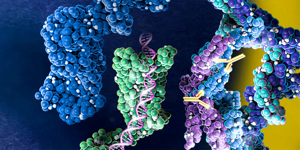
A new article in Nature Scientific Reports answers open questions about TOPBP1, a protein involved in repairing DNA double-strand breaks (DSBs). The study used cell-free protein expression and a unique DSB system to identify domains that were important for activation of a protein kinase.

Continue reading “Characterizing DNA Repair Proteins with Cell-Free Protein Expression”

Looking Back: Cell-Free Expression Systems Helped to Characterize Proteins Involved in Hypoxia Response

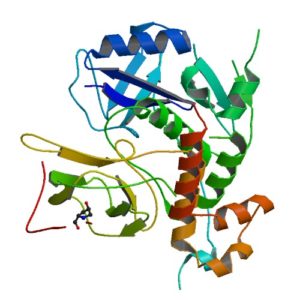
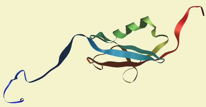
Structur of a HIF-1a-pVHL-ElonginB-ElonginC complex
Structure of a HIF-1a-pVHL-ElonginB-ElonginC complex

William G. Kaelin Jr., Sir Peter J. Ratcliffe and Gregg L. Semenza were awarded the 2019 Nobel Prize in Physiology or Medicine for their discoveries of how cells sense and adapt to oxygen availability.

Kaelin and Ratcliffe’s labs focused their efforts on the transcription factor HIF (hypoxia-inducible factor). This transcription factor is critical in the cellular adaptation of to changes in oxygen availability.

When oxygen levels are elevated cells contain very little HIF. Ubiquitin is added to the HIF protein via the VHL complex and it is degraded in the proteasome.  When oxygen levels are low (hypoxia) the amount of HIF increases.

In 2001 both groups published articles characterizing the interaction between VHL and HIF, and these articles were referenced by the Nobel Prize Organization in their press release about this year’s award. (1,2). Both studies demonstrated that under the normal oxygen conditions hydroxylation of proline residue P564 enabled VHL to recognize and bind to HIF.

The use of cell free expression (i.e., TNT Coupled Transcription/Translation System) by both labs was key in the characterization of the VHL:HIF interaction The labs utilized HIF and VHL 35-S labeled proteins generated via the TNT system under both normal or in a hypoxic work station to:

  • Determine the affect of ferrous chloride and cobaltous chloride on the interaction
  • Map the specific region of HIF required for the interaction to occur (556-574)
  • Determine the effect of HIF point mutations on the interaction
  • Use synthetic peptides to block the interaction
  • Conclude that a factor in mammalian cells was necessary for the interaction to occur.

Literature Cited

  1. Ivan, M et al. (2001) HIF Targeted for VHL-Mediated Destruction by Proline Hydroxylation: Implications for O2 Sensing. Science 292: 464–67.
  2. Jaakkola, P. et al. (2001) Targeting of HIF-α to the von Hippel-Lindau Ubiquitylation of Complex by O2– Regulated Prolyl Hydroxylation. Science 202, 468–72 .

Related Posts

Luciferase Immunoprecipitation System Assay (LIPS): Expression of Luciferase Antigen using TNT Transcription/Translation Kit

NanoLuc dual reporters
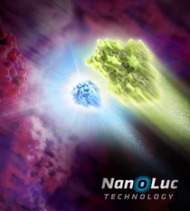
Illustration showing NanoLuc and firefly luciferase reporters.

The luciferase immunoprecipitation system (LIPS) assay is a liquid phase immunoassay allowing high-throughput serological screening of antigen-specific antibodies. The immunoassay involves quantitating serum antibodies by measuring luminescence emitted by the reporter enzyme Renilla luciferase (Rluc) fused to an antigen of interest. The Rluc-antigen fusion protein is recognized by antigen-specific antibodies, and antigen-antibody complexes are captured by protein A/G beads that recognize the Fc region of the IgG antibody (1).

In a recent publication (2), this assay was used to assess the presence of autoantibodies against ATP4A and ATP4B subunits of parietal cells H+, K+-ATPase in patients with atrophic body gastritis and in controls. Continue reading “Luciferase Immunoprecipitation System Assay (LIPS): Expression of Luciferase Antigen using TNT Transcription/Translation Kit”

Rabbit Reticulocyte Lysate Translation Systems: Tools for the analysis of translational regulation

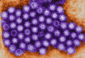
TEM of Norovirus particles. Photo Credit: Charles D. Humphrey, Centers for Disease Control and Prevention

Rabbit Reticulocyte Lysate Translation Systems are used in the identification of mRNA species, the characterization of their protein products and the investigation of transcriptional and translational control. Rabbit Reticulocyte Lysate is prepared from New Zealand white rabbits. After the reticulocytes are lysed, the extract is treated with micrococcal nuclease to destroy endogenous mRNA and thus reduce background translation to a minimum.

Untreated Lysate is prepared from New Zealand white rabbits in the same manner as treated lysates with the exception that it is not treated with micrococcal nuclease. Unlike a coupled system that initiates transcription/translation from DNA, the RNA-based rabbit reticulocyte can be used for the direct investigation of transcriptional/translational control and the replication of RNA-based viruses.


References

Characterization of translation regulation (i.e., UTRs, Capping, IRES)

  1. Nguyen, H-L .et al. (2013) Expression of a novel mRNA transcript for human microsomal epoxide hydrolase is regulated by short reading frames within it 5’ –untranslated region. RNA. 19, 752–66.
  2. Wei, J. et al. (2013) The stringency of start codon selection in the filamentous fungus Neurospora crass. J. Biol. Chem. 288, 9549–62.
  3. Paek Ki-Y. et al. (2012) Cap-Dependent translation without base-by-base scanning of an messenger ribonucleic acid. Nucl. Acid. Res. 40, 7541–51.
  4. Se, and NH. Su.W. et al. (2011) Translation, stability, and resistance to decapping of mRNA containing caps substituted in the triphosphate with BH3. RNA 17, 978–88.
  5. Anderson, D. et al. (2011) Nucleoside modifications in RNA limit activation of 2’-5’ oligoadenylate synthetase and increase resistance to cleavage by RNase L. Nucl. Acid. Res. 39, 9329-38.

RNA virus Characterization

  1. Vashist, S. et al. (2012) Identification of RNA-protein interaction networks involved in the Norovirus life cycle. J. Vir. 86, 11977–90.
  2. Soto-Rifo, R. et al. (2012) Different effects of the TAR structure on HIV-1 and HIV-2 genomics RNA translation. Nucl. Acids. Res. 40, 2653–67.
  3. Poyry, T. et al. (2011) Mechanisms governing the selection of translation initiation sites on Foot-and-Mouth Disease Virus RNA. J.Vir. 85, 10178–88.
  4. Cheng, E. et al. (2011) Characterization of the interaction between Hantavirus nucleopcapsid protein and ribosomal protein S19. J. Biol. Chem. 286, 11814–24.
  5. Vera-Otarola, J. et al. (2011) The Andes Hantavirus NSs Protein is expressed from the Viral mRMA by a leaky scanning mechanism. J. Vir. 86, 2176–87.

Cell free application: Sumoylation characterization

Small Ubiquitin-like Modifier (or SUMO) proteins are a family of small proteins that are covalently attached to and detached from other proteins in cells to modify their function. SUMOylation is a post-translational modification involved in various cellular processes, such as nuclear-cytosolic transport,
transcriptional regulation, apoptosis, protein stability and response to stress.

In contrast to ubiquitin, SUMO is not used to tag proteins for degradation. Mature SUMO is produced when the last four amino acids of the C-terminus have been cleaved off to allow formation of an isopeptide bond between the C-terminal glycine residue of SUMO and an acceptor lysine on the target protein.

Cell free expression can be used to characterize sumoylation of proteins. Target proteins are expressed in a rabbit reticulocyte cell free system (supplemented with necessary addition components,). Proteins that have been modified can be analyzed by a shift in migration on polyacrylamide gels, when compared to control reactions.

The following references illustrate the use of cell free expression for this application.

Brandl, A. et al. (2012) Dynamically regulated sumoylation of HDAC2 controls p53 deacetylation and restricts apoptosis following genotoxic stress. J. Mol. Cell. Biol. (online only)

Janer, A. et al. (2010). SUMOylation attenuates the aggregation propensity and cellular toxicity of the polyglutamine expanded ataxin-7. Human. Mol. Gen. 19, 181—95.

Rytinki, M. et al. (2009) SUMOylation attenuates the function of PGC-1alpha. J. Biol. Chem. 284, 26184-93.

Klein, U. et al. (2009) RanBP2 and SENP3 function in a mitotic SUMO2/3 conjugation-deconjugation cycle on Borealin. Mol. Cell. Biol. 20, 410–18.

Seo, W. and Ziltener, H. (2009) CD43 processing and nuclear translocation of CD43 cytoplasmic tail are required for cell homeostasis. Blood, 114, 3567–77.