NanoBiT™ Assay: Transformational Technology for Studying Protein Interactions Named a Top 10 Innovation of 2015

21416943-wb-cr-uw-nanobit-pm-page-hero
For three out of the last four years, we have been honored to have one of our key technologies named a Top 10 Innovation by The Scientist. This year the innovative NanoBiT™ Assay (NanoLuc® Binary Technology) received the recognition. NanoBiT™ is a structural complementation reporter based on NanoLuc® Luciferase, a small, bright luciferase derived from the deep sea shrimp Oplophorus gracilirostris.

Using plasmids that encode the NanoBiT complementation reporter, you can make fusion proteins to “report” on protein interactions that you are studying. One of the target proteins is fused to the 18kDa subunit; the other to the 11 amino acid subunit. The NanoBiT™ subunits are stable, exhibiting low self-affinity, but produce an ultra-bright signal upon association. So, if your target proteins interact, the two subunits are brought close enough to each other to associate and produce a luminescent signal. The strong signal and low background associated with a luminescent system, and the small size of the complementation reporter, all help the NanoBiT™ assay overcome the limitations associated with traditional methods for studying protein interactions.

The small size reduces the chances of steric interference with protein interactions. The ultra bright signal, means that even interactions among proteins present in very low amounts can be detected and quantified–without over-expressing large quantities of non-native fusion proteins and potentially disrupting the normal cellular environment. And the NanoBiT™ assay can be performed in real time, in live cells.

The NanoBiT™ assay is already being deployed in laboratories to help advance understanding of fundamental cell biology. You can see how one researcher is already taking full advantage of this innovative technology in the video embedded below:

Visit the Promega web site to see more examples more examples how the NanoBiT™ assay can break through the traditional limitations for studying protein interactions in cells.

You can read the Top 10 article in The Scientist here.

Biotechnology Ice Breakers: A Few Conversation Starters

quiz pictureThe biotechnology industry is one of the most dynamic out there – in fact, it never stands still! For non-scientists this can be intimidating. For scientists, it can be challenging to explain what we do in ways that non-scientists can understand and appreciate.

Scientists have made great strides in improving our ability to use molecular processes to our advantage, from discovering the basics of how to isolate and manipulate DNA to gaining an understanding of how genes direct the creation of proteins in cells.  It’s clear that there is a lot we can contribute to the scientific literacy of the general public.

In this spirit, we’ve designed a short quiz for both non-scientists (you may learn something new) and scientists (you may find it useful for engaging in conversations with your non-scientist friends and family members).  Spoiler alert: answers are provided. Continue reading “Biotechnology Ice Breakers: A Few Conversation Starters”

Reflecting on the Future: Hands-On, Person-to-Person Educational Experiences

iStock_000053412884LargeThe start of a new year is always a good time for reflection.  For those of us at the BioPharmaceutical Technology Center Institute (BTC Institute), this means looking at the programs we offer and considering ones we might like to develop.

In this process, we find ourselves continuing to feel certain that the hands-on, lab-based opportunities we provide add something meaningful to the education of those we serve, from middle school students and their teachers to graduate students to scientists in academia and industry.  The value of learning concepts and techniques in a well-equipped setting, working with teachers and volunteers who are dedicated scientists, is significant.

In addition to gaining an understanding of the basics of molecular biology so key to biotechnology, these programs are also designed to support the development of critical thinking skills so necessary to scientific literacy.

We think this is also the case for our scientific symposia (Wisconsin Stem Cell Symposium; Wisconsin Human Proteomics Symposium) and our International Forum on Consciousness.  These events enable attendees to interact with speakers and other participants in person – in an environment designed to encourage the exchange of information, ideas and perspectives. Continue reading “Reflecting on the Future: Hands-On, Person-to-Person Educational Experiences”

Mass Spectrometry Application: Antibody Quantitation for Preclinical PK studies

Isoform_Antibodies_LinkedInTherapeutic monoclonal antibodies (mAbs) represent the majority of therapeutics biologics now on the market, with more than 20 mAbs approved as drugs (1–3). During preclinical development of therapeutic antibodies, multiple variants of each antibody are assessed for pharmacokinetic (PK) characteristics across model systems such as rodents, beagles and  primates. Ligand-binding assays (LBA) are the standard technology used to perform the PK studies for mAb candidates (4). Ligand-binding assays (LBAs) are methods used  to detect and measure a macromolecular interaction between a ligand and a binding molecule. In LBAs, a therapeutic monoclonal antibody is considered to be the ligand, or analyte of interest, while the binding molecule is usually a target protein.

LBAs have certain well-documented limitations (5). Specific assay reagents are often not available early in a program. Interferences from endogenous proteins, antidrug antibodies, and soluble target ligands are potential complicating factors.

Liquid chromatography coupled to tandem mass spectrometry (LC–MS/MS)-based methods represent a viable and complementary addition to LBA for mAb quantification in biological matrixes. LC–MS/MS provides specificity, sensitivity, and multiplexing capability.

A recent reference (6) illustrates an automated method to perform LC–MS/MS-based quantitation, with IgG1 conserved peptides, a heavy isotope labeled mAb internal standard,and anti-human Fc enrichment. The method was applied to the pharmacokinetic study of a mAb dosed in cynomolgus monkey, and the results were compared with the immunoassay data. The interesting finding of the difference between ELISA and LC–MRM-MS data indicated that those two methods can provide complementary information regarding the drug’s PK profile.

Literature Cited

  1. Mao, T. et al. (2013) Top-Down Structural Analysis of an Intact Monoclonal Antibody by Electron Capture Dissociation-Fourier Transform Ion Cyclotron Resonance-Mass Spectrometry. Anal.Chem. 85, 4239–46.
  2. Weiner, L. M. et al. (2010) Monoclonal antibodies: versatile platforms for cancer immunotherapy. Nat. Rev. Immunol. 10, 317–27.
  3. Nelson, A. et al. (2010) Development trends for human monoclonal antibody therapeutics. Nat. Rev. Drug Discovery. 9, 767–74.
  4. DeSilva, B. et al. (2003) Recommendations for the Bioanalytical Method Validation of Ligand-Binding Assays to Support Pharmacokinetic Assessments of MacromoleculesPharm. Res. 20, 1885–00.
  5. Ezan, E.et al. (2009) Critical comparison of MS and immunoassays for the bioanalysis of therapeutic antibodiesBioanalysis 1, 1375–88.
  6. Zhang, Q. et al. (2014) Generic Automated Method for Liquid Chromatography–Multiple Reaction Monitoring Mass Spectrometry Based Monoclonal Antibody Quantitation for Preclinical Pharmacokinetic Studies. Anal.Chem. 86, 8776–84.

Cell free application: Sumoylation characterization

Small Ubiquitin-like Modifier (or SUMO) proteins are a family of small proteins that are covalently attached to and detached from other proteins in cells to modify their function. SUMOylation is a post-translational modification involved in various cellular processes, such as nuclear-cytosolic transport,
transcriptional regulation, apoptosis, protein stability and response to stress.

In contrast to ubiquitin, SUMO is not used to tag proteins for degradation. Mature SUMO is produced when the last four amino acids of the C-terminus have been cleaved off to allow formation of an isopeptide bond between the C-terminal glycine residue of SUMO and an acceptor lysine on the target protein.

Cell free expression can be used to characterize sumoylation of proteins. Target proteins are expressed in a rabbit reticulocyte cell free system (supplemented with necessary addition components,). Proteins that have been modified can be analyzed by a shift in migration on polyacrylamide gels, when compared to control reactions.

The following references illustrate the use of cell free expression for this application.

Brandl, A. et al. (2012) Dynamically regulated sumoylation of HDAC2 controls p53 deacetylation and restricts apoptosis following genotoxic stress. J. Mol. Cell. Biol. (online only)

Janer, A. et al. (2010). SUMOylation attenuates the aggregation propensity and cellular toxicity of the polyglutamine expanded ataxin-7. Human. Mol. Gen. 19, 181—95.

Rytinki, M. et al. (2009) SUMOylation attenuates the function of PGC-1alpha. J. Biol. Chem. 284, 26184-93.

Klein, U. et al. (2009) RanBP2 and SENP3 function in a mitotic SUMO2/3 conjugation-deconjugation cycle on Borealin. Mol. Cell. Biol. 20, 410–18.

Seo, W. and Ziltener, H. (2009) CD43 processing and nuclear translocation of CD43 cytoplasmic tail are required for cell homeostasis. Blood, 114, 3567–77.

A New Method that Marks Proteins for Destruction

The ability to manipulate genes and proteins and observe the effects of specific changes is a foundational aspect of molecular biology. From the first site-directed mutagenesis systems to the development of knockout mice and RNA interference, technologies for making targeted changes to specific proteins to eliminate their expression or alter their function have made tremendous contributions to scientific discovery.

A recent paper highlights a novel application of HaloTag technology to enable the targeted destruction of specific HaloTag fusion proteins in vivo. The paper, published online in the July issue of Nature Chemical Biology, details a promising new method with application for validation of potential drug targets by specific in vivo inhibition, and for studying the function of specific genes in organogenesis or disease development. Continue reading “A New Method that Marks Proteins for Destruction”

Describing Life and Death in the Cell

4621CALife is complicated. So is death. And when the cells in your multiwell plate die after compound treatment, it’s not enough to know that they died. You need to know how they died: apoptosis or necrosis? Or, have you really just reduced viability, rather than induced death? Is the cytotoxicity you see dose-dependent? If you look earlier during drug treatment of your cells, do you see markers of apoptosis? If you wait longer, do you observe necrosis? If you reduce the dosage of your test compound, is it still cytotoxic? Continue reading “Describing Life and Death in the Cell”
A Sensitive, Universal Kinase Assay Ideal for Use with Low-Turnover Enzymes

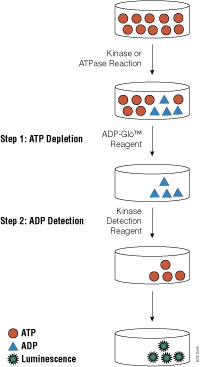
ADP-Glo™ Kinase Assay flowchart

Promega carries a large array of luminescent-based assays to measure cellular events such as viability, cytochrome P450 activity and apoptosis. Recently, we launched a new universal, homogeneous, high-throughput screening method called the ADP-Glo™ Kinase Assay, which measures kinase activity by quantifying the amount of ADP produced during a kinase reaction. While we already offer the Kinase-Glo™ Assays for assessing the quantity of ATP remaining after a kinase reaction, these assays are not ideal for use with low-activity kinases.

Continue reading “A Sensitive, Universal Kinase Assay Ideal for Use with Low-Turnover Enzymes”

Why Two Reporters are Better than One

As part of my job I occasionally search the literature for papers citing use of Promega products in new or interesting ways. Any search on dual-luciferase reporters usually generates a lot of returns. A search for dual-luciferase on Highwire press generates over 700 articles from 2009 alone. So why are dual-luciferase reporter assays so widely used? Continue reading “Why Two Reporters are Better than One”

Cell-Free Protein Synthesis

Cell-free protein synthesis (aka: in vitro translation) refers to protein production in vitro using lysates that provide the cellular machinery necessary for synthesis. Ribosomes, tRNAs, aminoacyl-tRNA synthetases, initiation/elongation/termination factors, GTP, ATP, Mg2+ and K+ are among the requirements for a translation system. These are provided by lysates, which can be from prokaryotic or eukaryotic sources, depending on your requirements.

Cell-free protein synthesis is most commonly used for generating protein for study of things like:

Continue reading “Cell-Free Protein Synthesis”