Three-dimensional (3D) cell culture systems have become essential tools in cancer research, drug screening and tissue engineering—offering a more physiologically relevant alternative to traditional 2D cultures, which often fail to replicate key in vivo microenvironment features. But as the field has evolved, variability in experimental outcomes has become a key challenge, limiting their reproducibility and translation into clinical settings. While spheroids offer layered architecture, nutrient gradients and multicellular interactions, inconsistent culture methods have made it difficult to draw reliable conclusions across labs.
At the time of writing this post, no scientist had yet discovered the secret to immortality. In our world, we’ve come to accept that living things are born, grow old and die—the circle of life.
And yet, for many years, life scientists believed that the circle of life did not apply to our constituent cells when cultured in a laboratory. That is, cultured normal human cells were immortal, and they would continue to grow and proliferate forever, as long as they were provided with the necessary nutrients.
Pioneering work published in 1961 by Leonard Hayflick and Paul Moorhead challenged that theory (reviewed in 1). Their research showed that normal cells in culture have a finite capacity to replicate. After they reach a certain number of replicative cycles, cells stop dividing. Hayflick and Moorhead made the important distinction between normal human cells and cultured cancer cells, which are truly immortal. In later years, the limit to the number of replicative cycles normal human cells can undergo became known as the Hayflick limit. Although some scientists still express skepticism about these findings, the Hayflick limit is widely recognized as a fundamental principle of cell biology.
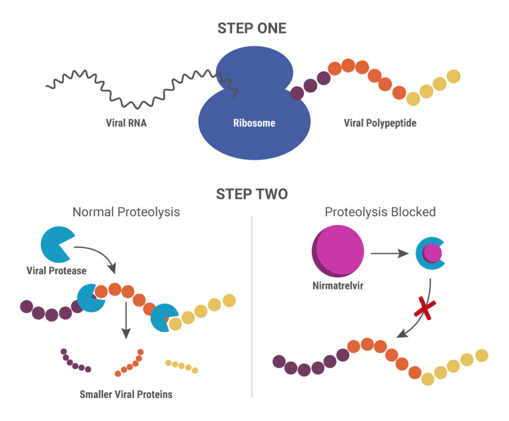
Cytochrome P450 (CYP) inhibitors are often used as boosting agents in combination with other drugs. This drug development strategy is front and center for Paxlovid, the new anti-SARS-CoV-2 treatment from Pfizer. Paxlovid is a combination therapy, comprised of two protease inhibitors, nirmatrelvir and ritonavir. It significantly reduces the risk of COVID-19 hospitalization in high-risk adults and is ingested orally rather than injected, which is an advantage over other SARS-CoV-2 treatments, such as Remdesivir.
Nirmatrelvir was originally developed by Pfizer almost 20 years ago to treat HIV and works by blocking enzymes that help viruses replicate. Pfizer created another version of this drug to combat SARS in 2003, but, once that outbreak ended, further development was put on pause until the advent of the COVID-19 pandemic. After developing an intravenous form of nirmatrelvir early in the pandemic, Pfizer created another version that can be taken orally and combined it with ritonavir.
When ritonavir was originally developed, it wasn’t considered particularly useful because it metabolized so quickly in the body. Now it is recognized as a pharmacokinetic enhancer in combination with other drugs. Ritonivir inhibits CYP3A4, an enzyme which plays a key role in the metabolism of drugs and xenobiotics. By inhibiting CYP3A4, ritonivir slows the metabolism of other drugs. In the case of Paxlovid, this allows nirmatrelvir to stay in the body longer at a high enough concentration to be effective against the virus. This ultimately means that patients can be given lower doses of the drug with reducing efficacy.
Nirmatrelvir inhibits the viral 3CL protease, so that functional, smaller viral proteins cannot be produced.
In 3D cell culture models, cells are grown under conditions that allow the formation of multicellular spheroids or microtissues. Instead of growing in a monolayer on a plate surface, cells in 3D culture grow within a support matrix that allows them to interact with each other, forming cell:cell connections and creating an environment that mimics the situation in the body more closely than traditional 2D systems. Although 3D cultures are designed to offer a more physiologically accurate environment, the added complexity of that environment can also present challenges to experimental design when performing cell-based assays. For example, it can be a challenge for assay reagents to penetrate to the center of larger microtissues and for lytic assays to disrupt all cells within the 3D system.
Earlier this week Terry Riss, a Senior Product Specialist at Promega, presented a Webinar on the challenges of performing cell-based assays on microtissues in 3D cell culture. During the Webinar, Terry gave an overview of the different methods available for 3D cell culture, providing a description of the advantages of each. He then discussed considerations for designing and optimizing cell-based assays for use in 3D culture systems, providing several recommendations to keep in mind when performing cell viability assays on larger microtissue samples.
This blog is written by guest author, Maggie Bach, Sr. Product Manager, Promega Corporation.
Researchers are increasingly relying on cells grown in three-dimensional (3D) structures to help answer their research questions. Monolayer, or 2D cell culture, was the go-to cell culture method for the past century. Now, the need to better represent in vivo conditions is driving the adoption of 3D cell culture models. Cells grown in 3D structures better mimic tissue-like structures, better exhibit differentiated cellular functions, and better predict in vivo responses to drug treatment.
Switching to 3D cell culture models comes with challenges. Methods to interrogate these models need to be adaptable and reliable for the many types of 3D models. Some of the most popular 3D models include spheroids grown in ultra-low attachment plates, and cells grown in an extracellular matrix, such as Matrigel® from Corning. Even more complex models include medium flow over the cells in microfluidic or organ-on-a-chip devices. Will an assay originally developed for cells grown in monolayer perform consistently with various 3D models? How is measuring a cellular marker different when cells are grown in 3D models compared to monolayer growth?
Snakebite is a serious public health issue in many tropical countries. Every year, roughly 2 million cases of poisoning from snakebites occur, and more than 100,000 people die. Snake venom is extremely complex, containing a cocktail of chemicals, many of which are undefined. This complicates the development of new therapeutics for treating snakebite.
Antivenom is the most effective treatment for snakebites,
but its production is complex and dangerous. It involves manually milking the
venom from different species of live snakes, then injecting small doses of the
venom into animals (mostly horses) to stimulate an immune response. After a
period of time, antibodies form in the animal’s blood, which is purified for
use as antivenom.
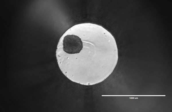
But what if we could produce snake venom in the lab, instead
of using live snakes? Recently, a group from the Netherlands did just that by
growing organoids derived from snake venom glands.
Traditionally, scientists have relied on flat,
two-dimensional cell cultures grown on substrates such as tissue culture
polystyrene (TCPS) to study cellular physiology. These models are simple and
cost-effective to culture and process. Within the last decade, however, three-dimensional
(3D) cell cultures have become increasingly popular because they are more
physiologically relevant and better represent in vivo conditions.
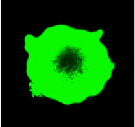
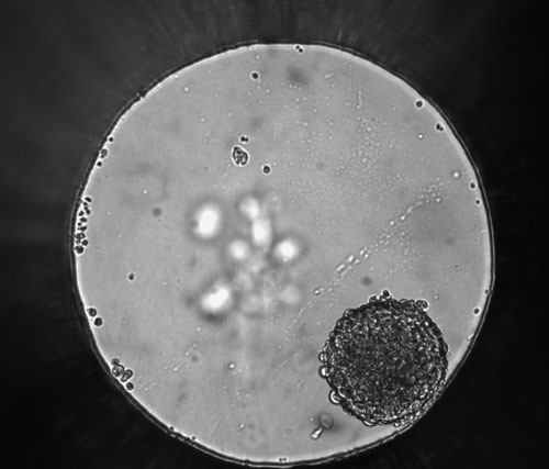
Differential contrast image of HCT116 colon cancer spheroid grown in a 96-well hanging-drop platform after seeding with 800 cells. Copyright Promega Corporation.Tissue culture using primary or cultured cell lines has long been a mainstay of testing compounds for inhibiting cell growth or promoting apoptosis during screening for cancer drugs. However, the standard culture conditions result in monolayers of cells, dividing and growing across the bottom of a well, plate or flask in a single layer. The drawback of this technique is that organisms do not come in monolayers; a three-dimensional (3D) spheroid is closer to the in vivo state, especially if the spheroids are made up of more than one cell type like tumors in multicellular organisms. Even more beneficial would be using 3D cultured cells in high-throughput screening to facilitate compound profiling for target effectiveness and cytotoxicity. In a recent PLOS ONE article, researchers used normal and breast cancer cells both in monoculture and coculture to test a set of compounds and found results differed between 2D and 3D cultured cells. Continue reading “Improving Cancer Drug Screening with 3D Cell Culture”
Based on the Illuminations article by Dr. Terry Riss, from our Cellular Analysis group.
Choosing the most appropriate cell health assay for your experiment can be difficult. There are several factors to consider when choosing an assay: the question you are asking, the nature of your sample, the number of samples being tested, the required sensitivity, the nature of the sample, the plates and plate readers and the reagent costs.
What question are you asking?
The first, and perhaps most important factor to consider, is the question you need answered. What do you want to know at the end of the experiment? There are cell health assays available that specifically detect the number of living cells, the number of dead cells, and for assessing stress response mechanisms or pathways that may lead to cell death. Matching the assay endpoint to the information you need is vital to choosing the appropriate cell health assay.
Search the PubMed database for “dual-luciferase” and citations abound. The Dual-Luciferase® Reporter Assay is a powerful tool that allows researchers to ask a multitude of questions about gene control and expression in a system that itself could be normalized and internally controlled. For more than 15 years, firefly and Renilla luciferases have formed the basis of a range of powerful assays and research tools for scientists who are asking questions about the deep and complex genetic and cellular story associated with cancer. Here we talk a bit of about bioluminescent chemistries, some of the newest bioluminescent tools available, and how some of these tools can be used to probe the deeper questions of cell biology, including cancer biology.
XWe use cookies and similar technologies to make our website work, run analytics, improve our website, and show you personalized content and advertising. Some of these cookies are essential for our website to work. For others, we won’t set them unless you accept them. To learn more about our approach to Privacy we invite you to Read More
By clicking “Accept All”, you consent to the use of ALL the cookies. However you may visit Cookie Settings to provide a controlled consent.
We use cookies and similar technologies to make our website work, run analytics, improve our website, and show you personalized content and advertising. Some of these cookies are essential for our website to work. For others, we won’t set them unless you accept them. To find out more about cookies and how to manage cookies, read our Cookie Policy.
If you are located in the EEA, the United Kingdom, or Switzerland, you can change your settings at any time by clicking Manage Cookie Consent in the footer of our website.
Necessary cookies are absolutely essential for the website to function properly. These cookies ensure basic functionalities and security features of the website, anonymously.
Cookie
Duration
Description
cookielawinfo-checbox-analytics
11 months
This cookie is set by GDPR Cookie Consent plugin. The cookie is used to store the user consent for the cookies in the category "Analytics".
cookielawinfo-checbox-functional
11 months
The cookie is set by GDPR cookie consent to record the user consent for the cookies in the category "Functional".
cookielawinfo-checbox-others
11 months
This cookie is set by GDPR Cookie Consent plugin. The cookie is used to store the user consent for the cookies in the category "Other.
cookielawinfo-checkbox-advertisement
1 year
The cookie is set by GDPR cookie consent to record the user consent for the cookies in the category "Advertisement".
cookielawinfo-checkbox-necessary
11 months
This cookie is set by GDPR Cookie Consent plugin. The cookies is used to store the user consent for the cookies in the category "Necessary".
cookielawinfo-checkbox-performance
11 months
This cookie is set by GDPR Cookie Consent plugin. The cookie is used to store the user consent for the cookies in the category "Performance".
gdpr_status
6 months 2 days
This cookie is set by the provider Media.net. This cookie is used to check the status whether the user has accepted the cookie consent box. It also helps in not showing the cookie consent box upon re-entry to the website.
lang
This cookie is used to store the language preferences of a user to serve up content in that stored language the next time user visit the website.
viewed_cookie_policy
11 months
The cookie is set by the GDPR Cookie Consent plugin and is used to store whether or not user has consented to the use of cookies. It does not store any personal data.
Analytical cookies are used to understand how visitors interact with the website. These cookies help provide information on metrics the number of visitors, bounce rate, traffic source, etc.
Cookie
Duration
Description
SC_ANALYTICS_GLOBAL_COOKIE
10 years
This cookie is associated with Sitecore content and personalization. This cookie is used to identify the repeat visit from a single user. Sitecore will send a persistent session cookie to the web client.
vuid
2 years
This domain of this cookie is owned by Vimeo. This cookie is used by vimeo to collect tracking information. It sets a unique ID to embed videos to the website.
WMF-Last-Access
1 month 18 hours 24 minutes
This cookie is used to calculate unique devices accessing the website.
_ga
2 years
This cookie is installed by Google Analytics. The cookie is used to calculate visitor, session, campaign data and keep track of site usage for the site's analytics report. The cookies store information anonymously and assign a randomly generated number to identify unique visitors.
_gid
1 day
This cookie is installed by Google Analytics. The cookie is used to store information of how visitors use a website and helps in creating an analytics report of how the website is doing. The data collected including the number visitors, the source where they have come from, and the pages visted in an anonymous form.
Advertisement cookies are used to provide visitors with relevant ads and marketing campaigns. These cookies track visitors across websites and collect information to provide customized ads.
Cookie
Duration
Description
IDE
1 year 24 days
Used by Google DoubleClick and stores information about how the user uses the website and any other advertisement before visiting the website. This is used to present users with ads that are relevant to them according to the user profile.
test_cookie
15 minutes
This cookie is set by doubleclick.net. The purpose of the cookie is to determine if the user's browser supports cookies.
VISITOR_INFO1_LIVE
5 months 27 days
This cookie is set by Youtube. Used to track the information of the embedded YouTube videos on a website.
Performance cookies are used to understand and analyze the key performance indexes of the website which helps in delivering a better user experience for the visitors.
Cookie
Duration
Description
YSC
session
This cookies is set by Youtube and is used to track the views of embedded videos.
_gat_UA-62336821-1
1 minute
This is a pattern type cookie set by Google Analytics, where the pattern element on the name contains the unique identity number of the account or website it relates to. It appears to be a variation of the _gat cookie which is used to limit the amount of data recorded by Google on high traffic volume websites.