In the field of cancer research, accurately measuring cell proliferation is crucial for assessing the efficacy of therapeutic agents. This is particularly difficult with CDK 4/6 inhibitors, which arrest cells in the G1 phase without stopping their growth. This continued growth can skew results from proliferation assays which rely on factors that naturally scale with cell growth. These include mitochondrial activity (ATP levels), total cell protein, or mRNA as measured through the PRISM molecular barcoding strategy. Even though these cells are not dividing, the increase in these measurements can misleadingly suggest active proliferation.
There is growing awareness among researchers of these challenges. A recent study highlights these limitations by demonstrating the discrepancies that arise when using metabolic assays to assess cell proliferation after treatment with drugs that induce cell cycle arrest. This blog post delves into the study’s implications and demonstrates how one of Promega’s latest developments is poised to address these challenges.
CDK 4/6 Inhibitors Demonstrate the Need for a New Proliferation Assay
The need for alternative approaches to study cell proliferation is emphasized in a recent publication evaluating the efficacy of CDK 4/6 inhibitors in cancer treatment (Foy et al., 2024). While CDK4/6 inhibitors effectively arrest the cell cycle, they do not prevent cells from continuing to grow in size. This growth results in increased mitochondrial activity and consequently, elevated ATP levels, complicating the use of assays that employ ATP as a proxy for cell number.
In their study, the authors treated a panel of eight cancer cell lines with 1 µM of the CDK 4/6 inhibitor palbociclib and evaluated cell proliferation using CellTiter-Glo® and CyQuant™, a DNA content-based assay. Treated cells arrest in the G1 phase of the cell cycle but continue to grow in size and increase their mitochondrial content. As a result, ATP levels increased while DNA content, as measured by CyQuant™ Direct, did not change. CellTiter-Glo® has been instrumental in evaluating cellular health, especially for massive efforts like the NCI-60 Screen; however, it must be recognized that other assays may be more suitable in situations similar to those/that described in the paper.
Addressing the Need for a New Method to Study Cell Proliferation
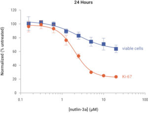
Foy and colleagues make clear the issues that can arise when using metabolism-based assays to assess cell proliferation; additionally, other common techniques require laborious wash steps or rely on indirect markers, radioactive material, and specialized equipment. To address these challenges, Promega developed the Lumit® hKi-67 Immunoassay for Cell Proliferation for detection of hKi-67, a well-known marker of cell proliferation. This assay increases confidence by tracking a well-known and metabolism independent marker of cell proliferation. It decreases lab time using a fast, no-wash, add-and-read protocol, and can provide an earlier readout of proliferation compared to CyQuant™ (Fig 1).


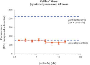
Fig 1. Treatment of HCT 116 cells (10,000/well, 96-well plate) with increasing concentrations of the antiproliferative agent nutlin-3A did not induce cytotoxicity and reduced Ki-67 expression levels in a dose-dependent manner. Ki-67 levels were assessed with the Lumit® hKi-67 Assay, while changes in viable cell number were determined with CyQUANT™ Direct in a parallel assay plate.
This assay utilizes Lumit® technology, which relies on labeled primary antibodies that in the presence of hKi-67, form a fully functional NanoBiT® luciferase enzyme. Addition of substrate results in a luminescent signal proportional to Ki-67 levels (Fig 2).

Fig 2. How the Lumit® hKi-67 Immunoassay Works
In conclusion, the publication discussed in this blog post highlights an opportunity for improvement; metabolic activity-based assays can indicate cell proliferation rather than cell growth alone. Promega’s innovative Lumit® hKi-67 Immunoassay for Cell Proliferation bridges this gap by tracking a marker that is independent of the metabolic state of cells, enabling researchers to be more confident in their assessment of proliferation. Through the expansion of its suite of cutting-edge tools, Promega continues to provide researchers with innovative solutions for understanding cell health.
Learn more about Promega’s Cell Health Portfolio.
Latest posts by Simon Moe (see all)
- Alzheimer Disease and Metabolic Dysfunction: A Critical Intersection in Brain Health - February 26, 2025
- Glo-ing Above and Beyond: Simplifying Science with MyGlo Reagent Reader - January 27, 2025
- The Greatness of Glycogen: A Central Storage Molecule in Energy Metabolism - January 3, 2025
