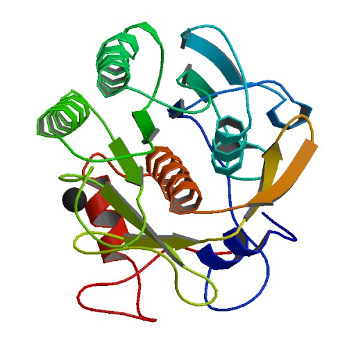
If you enter any molecular lab asking to borrow some Proteinase K, lab members are likely to answer: “I know we have it. Let me see where it is”. Sometimes the enzyme will be found to have expired. The lab may also have struggled with power outages or freezer malfunctions in the past. But the lab still decides to keep the enzyme. One may rightly ask – why do labs hang on to Proteinase K even when it has been stored under sub-standard conditions?
First of all Proteinase K is an amazing enzyme. It is stable under very harsh conditions. For example it is active at high temperature, under extreme pH, in the presence of SDS and EDTA; these are the conditions at which most of DNAses are inactivated (1). For that reason it is very useful for the isolation of native, undamaged DNAs or RNAs, since most microbial or mammalian DNases and RNases are rapidly chewed up by the enzyme, particularly in the presence of 0.5–1% SDS (2). It is the preferred method of nucleic acid isolation whenever high molecular weight genomic DNA is desired. In regards to temperature, raising the temperature of the reaction from 37°C to 50–60°C can increase the enzyme activity several fold. Heat treatment for 10–15 minutes at 65°C causes inhibition by less than 20–25%. Even when heated at 95°C for 10 minutes, Proteinase K still has some enzymatic activity. Based on these facts it is not hard to understand why occasional room temperature storage is considered harmless.
Did you know that you can order Proteinase K in solution or lyophilized Proteinase K from our website or even have it added to your local Helix unit for on-site stocking.
For all above mentioned characteristics, “ProK” is nowadays better known for its role in isolation of nucleic acids (3–6), than for protein fingerprinting applications (7). Current nucleic acid isolation kits and automated methods are mainly based on chaotropic salts, while old fashioned manual methods are based on Proteinase K. In such methods (6), Proteinase K alone in presence of NaCl2, SDS and EDTA is used to isolate highly pure, high molecular weight DNA. Usually researchers incubate minced tissue (e.g. mouse tail) overnight at 56°C with Proteinase K. The following day only a cloudy liquid is present where once there was intact tissue. If SDS and EDTA were not added that liquid would consist mainly of cells. Proteinase K activity is not localized only to proteins connecting the cells. Some membranes might also be damaged because the tissue was frozen or SDS was added to the ProK buffer. In both instances the bilipid layer of membranes will open and allow Proteinase K to digest whatever it encounters including nucleases. In the majority of methods, nucleases are additionally inactivated by the presence of EDTA, a chelating agent that removes Ca2+ and Mg2+ ions both of which are required for nuclease activity. The inactivation of nucleases is so efficient that some methods even allow tissue samples to be stored and preserved in Proteinase K without freezing until DNA is extracted. Unfortunately these are all laborious methods that take too much time. Research labs processing hundreds of samples a day need fast and automated methods. As mentioned earlier these methods are based on chaotropic salts that quickly denature proteins while allowing binding of nucleic acids to a charged surface of silica or cellulose. However, even these fast and automated protocols can benefit from pretreatment with Proteinase K. Here is how.
Are you looking for proteases to use in your research?
Explore our portfolio of proteases today.
Blood, serum, mucus, formalin-fixed paraffin-embedded (FFPE) tissue, hair, bones, mouse tail, and other sample types are full of proteins and other inhibitors which can interfere with the isolation of nucleic acids or downstream applications. The excess of proteins can prevent nucleic acid binding to a charged surface. They will render the lysate viscous, blocking movement of molecules, prevent nucleic acid binding to paramagnetic particles or clog the resin or the membrane. When proteins are denatured and chopped with Proteinase K prior to binding of nucleic acids there is significant increase in yield and purity. That initial lysate is subjected to further lysis with various lytic buffers and subsequent protein precipitation. These lytic buffers are not an ideal environment for proteinase K and will inactivate it quickly. That is the reason why we mix Proteinase K with a sample prior to addition of the lysis buffer. Even short sample pre-treatment with ProK significantly increases yield and purity of nucleic acids.
Concerned about the sustainability of products that you use a lot like Proteinase K? Check out our Green Sheet for Proteinase K.
Proteinase K pretreatment can be as long as overnight or as short as 15 minutes (9). Samples can be incubated with ProK in its traditional buffer with EDTA, SDS and NaCl2 or ProK can be mixed directly with samples just prior to addition of lysis buffer containing chaotropic salt. It really depends on the protocol developed by a kit manufacturer or on lab practices. If you choose to combine a traditional buffer with a modern kit, you better make sure that the ProK buffer composition will not interfere with subsequent steps used in the particular kit and method.
- Ebeling W, Hennrich N, Klockow M, Metz H, Orth HD, Lang H (1974). Proteinase K from Tritirachium album Limber. Eur. J. Biochem. 47 (1): 91–97.
- Hilz H., Wiegers, U. and Adameitz, P. (1975) Stimulation of Proteinase K Action by Denaturing Agents: Application to the Isolation of Nucleic Acids and the Degradation of ‘Masked’ Proteins Eur. J. Biochem. 56, 103–8
- Schwartz, D.C. and Cantor, C.R. (1984) Separation of yeast chromosome-sized DNAs by pulsed field gradient gel electrophoresis. Cell 37(1):67–75.
- Herrmann, B.G. and Frischauf, A.M. (1987) Isolation of genomic DNA. Meth.Enzymol. 152, 180–3.
- Lee, J.J. and Costlow, N.A. (1987) A molecular titration assay to measure transcript prevalence levels. Meth. Enzymol. 152, 633–48.
- Sambrook, J., Fritsch, E.F. and Maniatis, T. (1989) Molecular Cloning: A Laboratory Manual, Volume 3, Cold Spring Harbor Laboratory, Cold Spring Harbor, NY, B.16.
- Cleveland, D.W. et al. (1977) Peptide mapping by limited proteolysis in sodium dodecyl sulfate and analysis by gel electrophoresis. J. Biol. Chem. 252, 1102–6.
- Parzer, S. and Mannhalter, C. (1991) A rapid method for the isolation of genomic DNA from citrated whole blood. Biochem J. 273(Pt 1): 229–231.
Nives Kovacevic
Latest posts by Nives Kovacevic (see all)
- Practical Tips for HEK293 Cell Culture When Using cAMP-Glo™ Assay - January 31, 2014
- ProK: An Old ‘Pro’ That is Still In The Game - March 6, 2013
