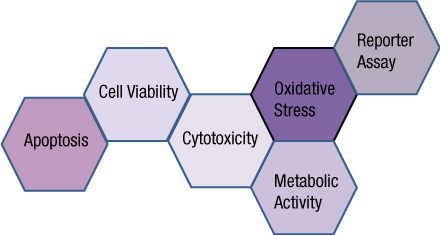
You often need several pieces of information to really understand what is happening within a cell or population of cells. If your cells are not proliferating, are they dying? Or, are you seeing cytostasis? If they are dying, what is the mechanism? Is it apoptosis or necrosis? If you are seeing apoptosis, what is the pathway: intrinsic or extrinsic?
If you are measuring expression of a reporter gene and you see a decrease in expression, is that decrease due to transfection inefficiencies, cytotoxicity, or true down regulation of your reporter gene?
To investigate these multiple parameters, you can run assays in parallel, but that requires more sample, and sample isn’t always abundant.
Multiplexing assays allows you to obtain information about multiple parameters or events (e.g., reporter gene expression and cell viability; caspase-3 activity and cell viability) from a single sample. Multiplexing saves sample, saves time and gives you a more complete picture of the biology that is happening with your experimental sample.

Multiplexing assay reagents to measure biomarkers in the same sample has often been considered an application only accomplished with antibodies or dyes and sophisticated detection instrumentation. However, Promega has developed microwell plate based assays for cells in culture that allow multiplexed detection of biomarkers in the same sample well using standard multimode multiwell plate readers.
A Better Understanding
A key benefit of multiplexing is a better understanding of the event being measured in the context of a second parameter. For example, multiplexing a cytotoxicity assay with a viability assay can allow differentiation of cytotoxicity from cytostasis, normalization of cell number well to well, or reveal assay interferences. Multiplexing different biomarkers of toxicity can allow you to determine the mechanism of toxicity or the appropriate timing to apply a confirmatory assay. For example, performing a real-time cytotoxicity assay followed by a caspase assay can allow you to determine the optimal window of caspase activation and confirm that the mechanism of toxicity is apoptosis. The table below presents some common objectives of multiplex experiments and example assay combinations that can be used to address them.
Perhaps the most common form of multiplexing practiced in the life sciences is the dual luciferase reporter assay, where one reporter is used to measure the experimental effect and a second reporter is used to measure the transfection efficiency or “background” of transcription/translation of an exogenous reporter. An easy yet robust dual-reporter assay consists of a firefly-based experimental reporter and a second Renilla-based normalization reporter. In situations where using a single genetic reporter is desirable, the reporter assay can be multiplexed with viability or cytotoxicity assays to differentiate changes in reporter gene expression from cytotoxic events.
Things to Consider
When considering multiplexing two or more assays, the assays must meet the following criteria:
- The signals for the multiple assays must be spectrally or temporally distinct.
- The assay chemistries must be compatible.
- The assays must fit into the same well or be easily separated (e.g., one assay uses the cells, the other uses the culture medium).
If these criteria are met, multiplexing can give you greater confidence in the parameter that you are measuring by providing context relative to other events happening in the cell or experimental system. Multiplexing cell-based assays with reporter assays, biochemical assays or other cell-based assays helps to normalize data, reduce work and materials, and lessen the variability associated with performing multiple replicates.
| Experimental Objective | Type of Multiplex | Example Assay 1 | Example Assay 2 |
| Normalize Reporter Activity to Transfection Efficiency or Viability | Dual-reporter assay | Firefly reporter detection reagent | Renilla reporter detection reagent |
| Reporter assay and viability assay | CellTiter-Fluor™ Cell Viablity Assay | One-Glo™ Luciferase Reporter Assay | |
| Distinguish Cytotoxicity from Cytostasis | Cytotoxicity assay /viability assay | CytoTox- Fluor™ Cytotoxicity Assay | CellTiter-Glo® Luminescent Cell Viability Assay |
| Differentiate Apoptosis from Necrosis | Cytotoxicity or viability assay/ Caspase assay | CellTox™Green Cytotoxicity Assay | Caspase-Glo® 3/7 Assay |
| Determine the Mechanism of Apoptosis | Two caspase assays | Apo-ONE® Homogeneous Caspase-3/7 Assay | Caspase-Glo® 8 or 9 Assay |
| Differentiate Mitochondrial toxicity from other cytotoxic events | cytotoxicity assay/viability assay (Available as Mitochondrial ToxGlo™ Assay) | Membrane integrity | ATP changes |
Michele Arduengo
Latest posts by Michele Arduengo (see all)
- An Unexpected Role for RNA Methylation in Mitosis Leads to New Understanding of Neurodevelopmental Disorders - March 27, 2025
- Unlocking the Secrets of ADP-Ribosylation with Arg-C Ultra Protease, a Key Enzyme for Studying Ester-Linked Protein Modifications - November 13, 2024
- Exploring the Respiratory Virus Landscape: Pre-Pandemic Data and Pandemic Preparedness - October 29, 2024
