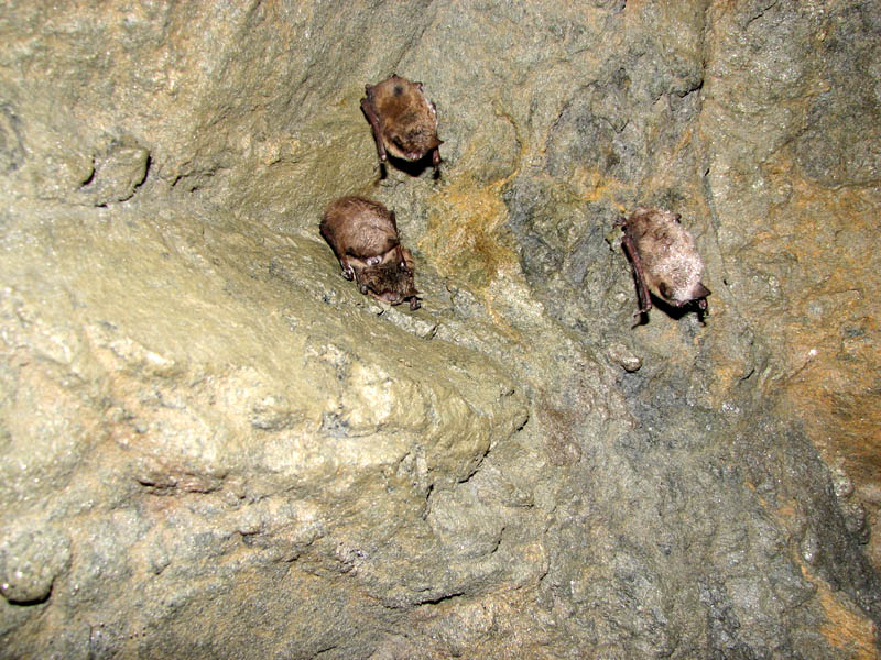
Since the first photograph of bats with white muzzles in Albany, NY, was published, hibernating bat populations in the northeastern U.S. have been devastated by an emerging disease, White-Nose Syndrome (WNS), which continues to spread throughout the United States and now has been found in two Canadian provinces. Bats suffering from WNS are emaciated with little or no body fat and have a characteristic white fungal growth on their wing membranes, ears and muzzles. Instead of hibernating all winter, these bats can be seen active in the snow, when there is virtually no food available.
The infectious agent that responsible for WNS is a new species of the cold-loving fungus, Pseudogymnoascus destructans (formerly, Geomyces destructans) . Currently, WNS is confirmed either through histological analysis or by fungal culture. Both of these techniques have significant limitations. First, they have turnaround times of at least one week. Secondly, they require large amounts of tissue sample from affected bats, more than can be reasonably taken from live bats. Histological analysis is a laborious process that requires highly skilled and trained personnel. Fungal culture can be difficult because bats harbor many bacteria and fungi, and getting a pure culture of a causative organism is not simple. Furthermore, researchers need a quick method for assessing spread of the disease that can provide results quickly.
Polymerase Chain Reaction (PCR), already used for a host of diagnostic tests in humans, plants and animals, is a logical choice. In a paper published in the Journal of Veterinary Diagnostic Investigation, Lorch and colleagues design and evaluate a PCR-based diagnostic method for WNS. They compare the PCR method to a fungal culture method and the “gold standard” traditional histological analysis.
For the culture method, researchers obtained 1.5 × 1.5cm samples of wing tissue from bats carcasses submitted to the U.S. Geological Survey–National Wildlife Health Center in Madison, WI. These samples were cultured in medium plus antibiotic for 10–30 days, and checked every one 1–3 days for the distinctively shaped conidia that are hallmarks of G. destructans colonies. To confirm isolation of G. destructans, the internal transcribed spacer (ITS) region of the rRNA genes was sequenced.
For the PCR-based diagnostic method, genomic DNA was isolated from a 3 × 3mm plug of wing tissue obtained next to the sample used for the culture method. Genomic DNA was isolated using a modification of a commercially available protocol. Two forward and three reverse primers were synthesized spanning the small subunit (18S) rRNA gene sequences. The pair giving the strongest and most consistent PCR product was chosen for the diagnostic applications.
Of the 78 carcasses examined in this study, 48 met the histological criteria for WNS. Using the culture technique, researchers were only able to isolate G. destructans from 26 of the 78 carcasses, giving a diagnostic sensitivity for the culture method of only 54%. No false positives were obtained using the culture technique. Using the PCR method, detected G. destructans DNA in 46 of the 78 carcasses, giving a 96% diagnostic sensitivity. No false positives were detected, making this assay highly specific for detecting G. destructans.
Clearly the PCR method performed well and is more timely and less labor-intensive than the culture method for detecting G. destructans. It is not as sensitive as the histological method, but it uses far less tissue, requires less time, and less skill and training. These advantages provide a tool that will allow researchers to collect larger numbers of samples over time to monitor and track WNS and hopefully, to eventually assess effectiveness of any treatments for WNS as they are developed.
![]() Lorch, J.M., Gargas, A, Meteyer C.U., Berlowski-Zier, B.M., Green, D.E., Shearn-Bochsler, V., Thomas N.J., & Blehert, D.S. (2010). Rapid polymerase chain reaction diagnosis of white-nose syndrome in bats. Journal of veterinary diagnostic investigation : official publication of the American Association of Veterinary Laboratory Diagnosticians, Inc, 22, 224–30 PMID: 20224080
Lorch, J.M., Gargas, A, Meteyer C.U., Berlowski-Zier, B.M., Green, D.E., Shearn-Bochsler, V., Thomas N.J., & Blehert, D.S. (2010). Rapid polymerase chain reaction diagnosis of white-nose syndrome in bats. Journal of veterinary diagnostic investigation : official publication of the American Association of Veterinary Laboratory Diagnosticians, Inc, 22, 224–30 PMID: 20224080
Michele Arduengo
Latest posts by Michele Arduengo (see all)
- An Unexpected Role for RNA Methylation in Mitosis Leads to New Understanding of Neurodevelopmental Disorders - March 27, 2025
- Unlocking the Secrets of ADP-Ribosylation with Arg-C Ultra Protease, a Key Enzyme for Studying Ester-Linked Protein Modifications - November 13, 2024
- Exploring the Respiratory Virus Landscape: Pre-Pandemic Data and Pandemic Preparedness - October 29, 2024

I think you should have used “…wins by a nose…” in the title.