Striking Fear into the Heart of Cardiovascular Disease Using Zebrafish and NanoLuc® Luciferase

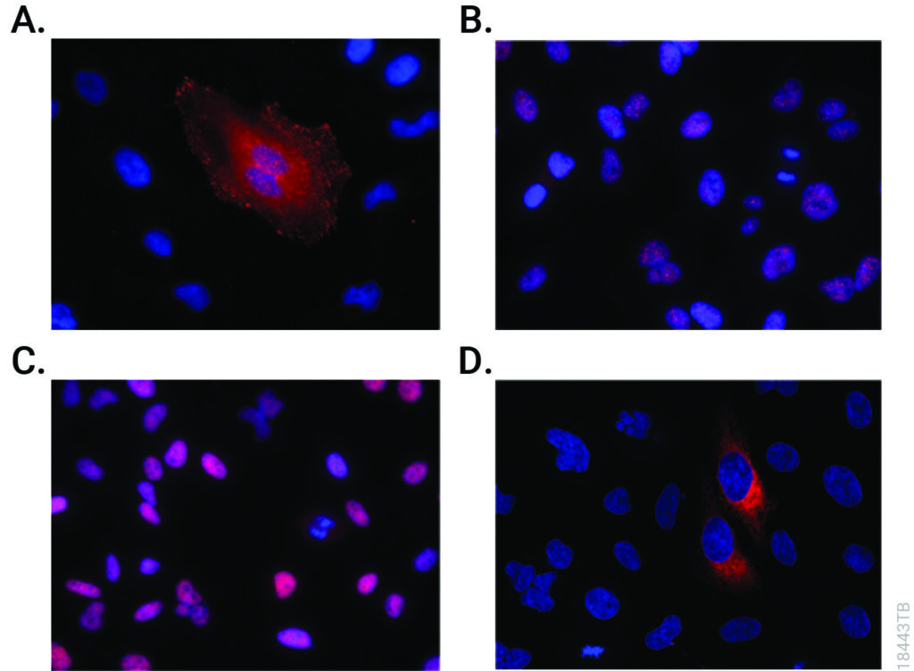
Representative images of ApoB-LP localization in zebrafish across developmental, genetic, pharmacological and dietary manipulations.
Credit: Figure 5.D of The LipoGlo reporter system for sensitive and specific monitoring of atherogenic lipoproteins by James Thierer, Stephen C. Ekker and Steven A. Farber.
Article licensed under Creative Commons Attribution 4.0 International License.

Cardiovascular diseases, or CVDs, are collectively the most notorious gang of cold-blooded killers threatening human lives today. These unforgiving villains, including the likes of coronary heart disease, cerebrovascular disease and pulmonary embolisms, are jointly responsible for more deaths per year than any other source, securing their seat as the number one cause of human mortality on a global scale.

One of the trademarks of most CVDs is the thickening and stiffening of the arteries, a condition known as atherosclerosis. Atherosclerosis is characterized by the accumulation of cholesterol, fats and other substances, which together form plaques in and on the artery walls. These plaques clog or narrow your arteries until they completely block the flow of blood, and can no longer supply sufficient blood to your tissues and organs. Or the plaques can burst, setting off a disastrous chain reaction that begins with a blood clot, and often ends with a heart attack or stroke.

Given the global prevalence and magnitude of this problem, there is a significant and urgent demand for better ways to treat CVDs. In a recent study published in Nature Communications, researchers at the Carnegie Institution for Science, Johns Hopkins University and Mayo Clinic are taking the fight to CVDs through the study of low-density lipoproteins (LDLs), the particles responsible for shuttling bad cholesterol throughout the bloodstream.

Continue reading “Striking Fear into the Heart of Cardiovascular Disease Using Zebrafish and NanoLuc® Luciferase”

I Have My Luciferase Vector, Now What?

Choosing and Optimizing Transfection Methods

Here in Technical Services we often talk with researchers at the beginning of their project about how to carefully design and get started with their experiments. It is exciting when you have selected the Luciferase Reporter Vector(s) that will best suit your needs; you are going to make luminescent cells! But, how do you pick the best way to get the vector into your cells to express the reporter? What transfection reagent/method will work best for your cell type and experiment? Do you want to do transient (short-term) transfections, or are you going to establish a stable cell line?

Continue reading “I Have My Luciferase Vector, Now What?”

Manipulating Koala Microbiomes Using…Poo Pills?

“Koalas – mum and baby” by Amanda Penrose is licensed under CC BY-NC-SA 2.0 

In recent years, it’s become a well-documented fact that koalas are about as picky as they are adorable. These beloved Australian marsupials have evolved to become ecological specialists: consumers that feed primarily on a single organism, or small number of organisms. Eucalyptus, their organism of choice, encompasses approximately 900 species, most of which are native to Australia. To the koala’s benefit, the leaves of eucalyptus trees are difficult to digest, low in protein content and their chemical composition contains compounds that are toxic. This makes their competition for eucalyptus with other species virtually nonexistent.

That’s not to say there isn’t competition amongst themselves. Of those 900 species of eucalyptus, koalas are only really known to feed on about 40–50 of them, and of those 40–50, they tend to limit their diet to around 10. Depending on their location, however, some koalas will only stick to one preferred type, which can lead to trouble.  

Continue reading “Manipulating Koala Microbiomes Using…Poo Pills?”

Prions Go Slow with ASOs: Experimental Treatment for ALS, Alzheimer’s and Other Prion-like Diseases

In the late-80’s through the 90’s, food and health agencies focused on a mysterious fatal brain disease that infected thousands of cattle. Bovine spongiform encephalitis—or “mad cow disease”—is caused by an infectious protein called a prion. Despite fears that tainted meat would cause the disease to spread to humans, mad cow disease never really made an impact on human health. However, forms of the prion disease such as Creutzfeldt-Jakob disease do affect humans.

In addition to Creutzfeldt-Jakob disease, many neurodegenerative diseases such as Alzheimer’s, Parkinson’s, Huntington’s and amyotrophic lateral sclerosis (ALS or Lou Gehrig’s disease) are now thought to be a result of prion-like activity. There is no cure for these diseases, however, new experimental treatment strategies might help slow the progression of neural degeneration.

prion_bse_histology
The tell-tale “holes” of prion infection in brain tissue.
Continue reading “Prions Go Slow with ASOs: Experimental Treatment for ALS, Alzheimer’s and Other Prion-like Diseases”

Anti-Cancer Drugs Are Pro-Coral

With average sea surface temperatures increasing around the world, coral bleaching events are growing in extent and severity. More than two thirds of the corals in the Great Barrier Reef, the world’s largest coral reef, have already bleached. While the physiological consequences of coral bleaching are well-studied, we still don’t fully understand how bleaching happens on a cellular level.

Corals living on shallow patch reefs in Palau. The Palau International Coral Reef Center is the staging ground for the research on mechanisms allowing corals to thrive in warming waters.

Steve Palumbi at Stanford University is delving deeper into the mechanisms by which coral bleaching occurs. In 2018, Promega pledged $3 million over three years to the nonprofit Revive & Restore Catalyst Science Fund, to identify and develop advanced techniques for conservation, enhancing biodiversity, and genetic rescue. Palumbi was awarded the first grant from this fund to study the genomic stress trigger that causes corals to bleach in warming oceans.

Continue reading “Anti-Cancer Drugs Are Pro-Coral”

Delving into the Diversity of The Plague of Justinian

Wayson stain of Yersinia pestis showing the characteristic
Yersinia pestis. U.S. Center for Disease Control [Public domain], via Wikimedia Commons.
Human teeth play a key role in our understanding of how organisms evolve. Whenever a possible new member of the hominid family is uncovered, the shape and number of teeth are used to place that individual in the family tree. Teeth also harbor information about pathogens that have plagued humans for millennia. Because bacteria use our bloodstream as a transport system, protected places that can preserve DNA—like the pulp of teeth—are a rich medium for uncovering information about humans and the microbes that infected them.

Teeth have been the choice for identifying the infectious agent behind the Plague of Justinian in the sixth century and the Black Plague in the 14th century. In fact, Yersinia pestis, the bacterium responsible for these plagues, has infected humans as far back as the Neolithic. But what can we learn about the pandemic strain or strains of Y. pestis described in historical records? A team of researchers from Europe and the US, many of whom have been delving into the history of Y. pestis for the last decade, wanted to further investigate the Plague of Justinian. They studied bacterial DNA extracted from human remains found in Western European communal graves that were dated to around 541–750, the period of the historically documented Plague of Justinian. Their investigation examined the bacteria’s diversity and how far it spread during this “First Pandemic” of plague. Continue reading “Delving into the Diversity of The Plague of Justinian”

How Antioxidants Stimulate Lung Cancer Metastasis

You may have heard that antioxidants are good for you, but in some cases, they can be harmful. In 2014, a study led by Dr. Martin Bergo at the Karolinska Institutet in Sweden showed that antioxidant supplements, such as vitamin E, accelerated tumor growth. This sparked much controversy as it was previously believed that antioxidants prevented tumor progression.

Since then, more evidence suggests that antioxidants indeed promote tumor progression by reducing reactive oxygen species (ROS) that block tumor growth. In 2019, the same group published a follow-up study to further explain how antioxidants promote lung cancer metastasis.

Continue reading “How Antioxidants Stimulate Lung Cancer Metastasis”

Immunotherapy—Don’t Forget the Microbiome

Bacteria make you sick. The idea that bacteria cause illness has become ingrained in modern society, made evident by every sign requiring employees to wash their hands before leaving a restroom and the frequent food recalls resulting from pathogens like E. coli. But a parallel idea has also taken hold. As microbiome research continues to reveal the important role that bacteria play in human health, we’re starting to see the ways that the microbiota of the human body may be as important as our genes or environment.

The story of how our microbiome affects our health continues to get more complex. For example, researchers are now beginning to understand that the composition of bacteria residing in your body can significantly impact the effects of therapeutic drugs. This is a new factor for optimizing drug response, compared to other considerations such as diet, interaction with other drugs, administration time and comorbidity, which have been understood much longer.

Continue reading “Immunotherapy—Don’t Forget the Microbiome”

Kinase Inhibitors as Therapeutics: A Review

This blog was originally published in April of 2018. Today’s update includes the paper, “Quantitative, Wide-Spectrum Kinase Profiling in Live Cells for Assessing the Effect of Cellular ATP on Target Engagement” from Cell Chemical Biology, demonstrating the power of NanoBRET™ target engagement kinase assays in the study of kinase inhibitors.

The review “Kinase Inhibitors: the road ahead” was recently published in Nature Reviews Drug Discovery. In it, authors Fleur Ferguson and Nathanael Gray provide an up-to-date look at the “biological processes and disease areas that kinase-targeting small molecules are being developed against”. They note the related challenges and the strategies and technologies being used to efficiently generate highly-optimized kinase inhibitors.

This review describes the state of the art for kinase inhibitor therapeutics. To understand why kinase inhibitors are so important in the development of cancer (and other) therapeutics research, let’s start with the role of kinases in cellular physiology.

The road ahead for kinase inhibitor studies.

Why Kinases?

Continue reading “Kinase Inhibitors as Therapeutics: A Review”

Brazilian University Swatting at Leishmaniasis Parasite

The Medicinal Chemistry Center (CQMED), headquartered at Campinas State University in Brazil, recently started a project in partnership with Promega to develop drugs that can be used against Leishmania. This genus of protozoans is the etiological agent of leishmaniasis, transmitted to humans by sandflies.

Microscopic image of Leishmania parasite
Microscopic image of Leishmania tropica. Credit: Brian E. Keas at Michigan State University.

Leishmaniasis is classified as a neglected tropical disease that mainly affects poor communities. Symptoms include large skin sores and an enlarged spleen. The challenge in developing drugs to treat Leishmania is finding appropriate therapeutic targets. These targets are normally proteins whose inhibition leads to death of the parasite. In addition to pharmaceutical company Eurofarma, whose goal is to develop drugs for Leishmania, Promega was chosen to help solve this problem because of our NanoBRET™ Target Engagement (TE) assay*, a well-established technique for measuring protein interactions. In this assay, NanoLuc® luciferase is attached to the protein of interest, and a fluorescent NanoBRET™ tracer molecule is added to the cells. This produces a BRET signal. When a competing ligand is added, it will displace the tracer molecule, enabling quantification of the strength of the interaction compared to the tracer molecule..

A challenge that researchers will face will be ensuring that the NanoBRET™ tracer reaches the inside of the parasite cells; because Leishmania is an intracellular parasite, molecules need to cross the host cell membrane, the membrane of the vacuole containing the parasites, and the membrane of the parasite itself. Another challenge the slow reproduction of Leishmania within macrophages. On top of that is the fact that the parasite’s metabolism varies depending on its biological cycle, meaning that there could be long periods of time during which a drug’s therapeutic target is not expressed in the cell, during which time the drug would have no effect. The ideal target would be expressed at high levels throughout the cell cycle.

The project is being led by Rafael Couñago, a researcher at CQMED, and Promega scientists Matt Robers and Jean-Luc Vaillaud.

*An earlier version of this blog incorrectly said that these experiments are based on the NanoBRET™ assay using HaloTag® protein.