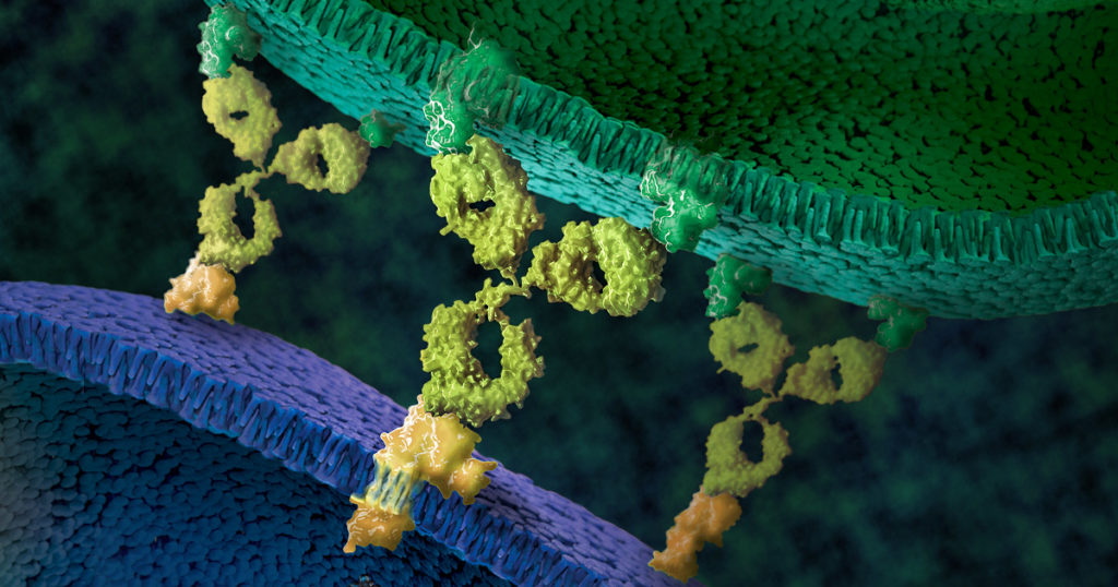
RIPK1: Promising Drug Target of Chronic Inflammatory Diseases

Today’s post is written by Michael Curtin, Senior Product Manager, Reporters and Signaling.

Inflammation is a defense mechanism that the body employs in which the immune system recognizes and removes harmful and foreign stimuli and begins the healing process. Inflammation can be either acute or chronic. Chronic inflammation is also referred to as slow, long-term inflammation and can last for prolonged periods (several months to years); chronic inflammation is caused by immune dysregulation. This typically takes the form of the body’s inability to resolve inflammation resulting from overproduction of inflammatory cytokines and chemokines, as well as danger-associated molecular patterns (DAMPs) released from dying cells (2). Tumor Necrosis Factor (TNF) is the primary cytokine involved in many common inflammatory diseases and is where many therapies targeting inflammation are focused.
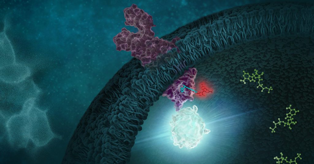
Signaling of kinases like RIPK1 can be studied using the NanoBRET target engagement assays

Recent research that RIP kinases (RIPK1 and RIPK3) are important regulators of innate immunity via their key roles in cell death signaling during cellular stress and following exposure to inflammatory and infectious stimuli. RIPK1 has an important scaffolding role in pro-inflammatory signaling where it interacts with TRADD, TRAF1 TRAF2, and TRAF3 and TRADD can act as an adaptor protein to recruit RIPK1 to the TNFR1 complex in a TNF-dependent process. RIPK1 plays a kinase activity-dependent role in both apoptotic and necroptotic cell death. A review article by Speir et al. (1) discusses the role of RIP kinases in chronic inflammation and the potential of RIPK1 inhibitors as a new therapeutic approach for the treatment of chronic inflammation. RIPK1 or Receptor Interacting Protein Kinase 1 is a serine/threonine kinase that was originally identified as interacting with the cytoplasmic domain of FAS. Promega offers several reagents that make studying RIPK1 easier- these include our RIPK1 Kinase Enzyme Systems which includes RIPK1 (Human, recombinant; amino acids 1-327), myelin basic protein (MBP) substrate, reaction buffer, MnCl2, and DTT and is optimized for use with our ADP-Glo Kinase Assay.

Continue reading “RIPK1: Promising Drug Target of Chronic Inflammatory Diseases”
Overcoming Challenges to Detect Apoptosis in 3D Cell Structures

This blog is written by guest author, Maggie Bach, Sr. Product Manager, Promega Corporation.

Researchers are increasingly relying on cells grown in three-dimensional (3D) structures to help answer their research questions. Monolayer, or 2D cell culture, was the go-to cell culture method for the past century. Now, the need to better represent in vivo conditions is driving the adoption of 3D cell culture models. Cells grown in 3D structures better mimic tissue-like structures, better exhibit differentiated cellular functions, and better predict in vivo responses to drug treatment.

Switching to 3D cell culture models comes with challenges. Methods to interrogate these models need to be adaptable and reliable for the many types of 3D models. Some of the most popular 3D models include spheroids grown in ultra-low attachment plates, and cells grown in an extracellular matrix, such as Matrigel® from Corning. Even more complex models include medium flow over the cells in microfluidic or organ-on-a-chip devices. Will an assay originally developed for cells grown in monolayer perform consistently with various 3D models? How is measuring a cellular marker different when cells are grown in 3D models compared to monolayer growth?

Close up of cells in 3D culture apparatus. 3D Cell Structures Provide Challenges for Measuring Markers of Cellular Activitiy
3D Cell Structures Provide Challenges for Measuring Markers of Cellular Activitiy
Continue reading “Overcoming Challenges to Detect Apoptosis in 3D Cell Structures”

World Firefly Day: Shining More Light on Glo-ing Innovations

On July 3rd and 4th, 2021, we celebrate World Firefly Day, and 2021, marks 30 years of luciferase products firefly luciferase vectors and Luciferase Assay System. These tools are key in advancing bioluminescent technology. To celebrate this day, we want to highlight some innovations that have been made possible with these tools.

Continue reading “World Firefly Day: Shining More Light on Glo-ing Innovations”

RAS-Targeted Drug Discovery: From Challenge to Opportunity

cancer cell, ras-targeted drug discovery

In 1963, Jennifer Harvey was studying Moloney murine leukemia virus (MMLV) at the cancer research department of the London Hospital Research Laboratories. After routine transfers of plasma from MMLV-infected rats to mice, she made an unusual discovery. In addition to the expected leukemia, the mice that received the plasma developed solid tumors (soft-tissue sarcomas), primarily in the spleen (1). A few years later, Werner Kirsten at the University of Chicago observed similar results working with mouse erythroblastosis virus (MEV) (2).

Subsequent research, with the advent of genome sequencing, showed that a cellular rat gene had been incorporated into the viral genome in both cases (3). These genomic sequences contained a mutation later shown to be responsible for the development of sarcomas, and the word “oncogene” became a common part of the vocabulary in cancer publications during the early 1980s (4). Harvey’s discovery led to the naming of the corresponding rat sarcoma oncogene as HRAS, while Kirsten’s related oncogene was named KRAS. Several laboratories, working independently, cloned the human homolog of the viral HRAS gene in 1982 (3). The human KRAS gene was cloned shortly thereafter, as well as a third RAS gene, named NRAS (3). Additional studies showed that a single point mutation in each of these genes led to oncogenic activation, and they have been popular targets for anticancer drug discovery efforts ever since.

Continue reading “RAS-Targeted Drug Discovery: From Challenge to Opportunity”

Antibody Response Differ in Adults, Kids and Potential Cross-Reactive Coronavirus Antibodies

B cell and B cell receptor cartoon. The B cell receptors are important for antibody response.
Drawing of a B cell and the B cell receptor. The receptor shows the characteristic Y shape of an immunoglobulin molecule.

B cells are the immune cells that produce antibodies (immunoglobulins or Ig) to detect intruding pathogens. B cells produce a variety of classes of antibodies. Generally during an immune response to a pathogen, whether viral or bacterial, B cells produce immunoglobulins (Ig) IgM and IgD, and later in the response, IgG and IgA, that are specific to the intruding organism. These Igs capture and aid in neutralizing the pathogen.

Ig classes can be studied by sequencing the B cell receptor (BCR), which binds antigen specifically. BCRs are formed via irreversible gene segment rearrangements of variable, diversity and joining (VDJ) genes. Ig classes can be diversified through somatic hypermutation and class-switch recombination of these gene segments (1).

B cell receptors with high sequence similarity can be found in individuals exposed to the same antigen, demonstrating that antigen exposure can result in similar B cell clones and memory B cells between individuals, both adults and children (1).

However, B cell immune responses can differ between adults and children. For example, children use more B cell clones that form neutralizing antibodies to HIV-1. And children infected with SARS-CoV-2 generally have milder illness than infected adults. SARS-CoV-2-infected children have lower antibody titers to the virus and more IgG-specific response to SARS-CoV-2 spike protein than to the nucleocapsid protein (1). These differences can contribute to faster SARS-CoV-2 clearance and lower viral loads in children versus adults.

Continue reading “Antibody Response Differ in Adults, Kids and Potential Cross-Reactive Coronavirus Antibodies”

Questions Arise about TMB as a Predictive Biomarker for Immune Checkpoint Inhibitor Therapy

Artist rendition of immune cells attacking a cancer cells. Immune checkpoint inhbitor therapy is a relatively new therapy for certain cancers.

Immune checkpoint inhibitor (ICI), or immune checkpoint blockade, therapies are a revolutionary, and relatively new, approach to treating cancer. These therapies work by blocking immune checkpoint proteins that act to negatively regulate the immune system through the PD-1 pathway. Some tumors express immune checkpoints to prevent the immune system from producing a strong enough immune response to kill the cancer cells. When these checkpoint proteins are blocked by an ICI, the body’s T-cells can recognize and kill the cancer cells. ICI therapies show tremendous promise. Unfortunately, not all tumors express immune checkpoint proteins, and so, not all tumors will be effectively treated with ICI therapies. The challenge is differentiating between the tumors that will respond and tumors that won’t.

DNA Mismatch Repair Deficiency Status as Detected by Microsatellite Instability or Immunohisotchemistry are Important Biomarkers for ICI

Biomarkers are measurable indicators of a clinical condition that can be found in tissue, blood, or other fluids. Predictive biomarkers for ICIs can help determine if these therapies are a suitable choice for treatment. Some tumors have deficiencies in their DNA mismatch repair mechanisms. Mismatch repair deficiency (dMMR) leads to the accumulation of mutations across the genome, particularly in microsatellites, which over time can result in higher levels of neoantigen production, rendering the tumors susceptible to the ICI therapy (1–5).

In 2017, Le et al. demonstrated that dMMR status reliably predicted response to an ICI therapy targeting the PD-1 checkpoint protein (6). Following this discovery, ICI based on dMMR  determined using either microsatellite instbility (MSI) or immunohistochemistry (IHC), gained clearance from the US Food and Drug Administation (FDA) for microsatellite instability-high (MSI-H) or dMMR by IHC solid tumors. This was the first time a cancer treatment was cleared based on a biomarker regardless of cancer origin (1,7).  Since then, MSI-H and dMMR, have become some of the most recognized tissue agnostic biomarkers for improved survival following ICI therapy of solid tumors (6,8,9).

Continue reading “Questions Arise about TMB as a Predictive Biomarker for Immune Checkpoint Inhibitor Therapy”

Bringing Cutting Edge Technologies to Academic Researchers Through the Academic Access Program

This post was written by guest blogger Iain Ronald, Director Academic/Government Market Segment at Promega.

My back story is similar to most of you reading this blog, high school education, undergraduate degree then onto a postgraduate degree. However, over 25 years ago during my undergraduate study, I was fortunate enough to work in the lab of Professor Ray Waters studying DNA damage in S. cerevisiae as a model organism and at the time PCR was cutting-edge technology and the PCR license was in full effect. However, there was one company that was fighting the good fight to democratize PCR for the good of the scientific community, Promega.

I became enamored with Promega then, and the next steps in my career were taken with a view to working at this company who, for all intents and purposes, seemed to really care about the progression of science beyond self-aggrandizement.

Now that I am working at Promega in a position where I can bring benefit to our academic community, I have pondered what I can do to equal the disruptive attitude I observed in this company all those years ago when they were fighting the then “big tech” for the enablement of the scientific community. 

Reporter bioassays are one of hte many offerings of the academic access program.
Continue reading “Bringing Cutting Edge Technologies to Academic Researchers Through the Academic Access Program”

How Promega Helped Our Lab Scale Up Drug Discovery for Bloodborne Pathogens

This blog was written by Sebastien Smick, Research Technician in Dr. Jacquin Niles’ laboratory at Massachusetts Institute of Technology (MIT)

Our lab is heavily focused on the basic biology and drug discovery of the human bloodborne pathogen Plasmodium falciparum, which causes malaria. We use the CRISPR/Cas9 system, paired with a TetR protein fused to a native translational repressor alongside a Renilla luciferase reporter gene, to conditionally knock down genes of interest to create modified parasites. We can then test all kinds of compounds as potential drug scaffolds against these gene-edited parasites. Our most recent endeavor, one made possible by Promega, is a medium-low throughput robotic screening pipeline which compares conditionally-activated or-repressed parasites against our dose-response drug libraries in a 384-well format. This process has been developed over the past few years and is a major upgrade for our lab in terms of data production. Our researchers are working very hard to generate new modified parasites to test. Our robots and plate readers rarely get a day’s rest!

Continue reading “How Promega Helped Our Lab Scale Up Drug Discovery for Bloodborne Pathogens”

Toxicity Studies in Organoid Models: Developing an Alternative to Animal Testing

Alternatives to animal testing have long been explored when it comes to studying the safety of various chemical compounds for use in food, medicine and cosmetics. With the advent of three-dimensional (3D) cell culture to create organoids, more relevant human organoid models are being explored as one way to provide safe and effective compound testing while minimizing the need for testing in animals. The international project Physiologically Anchored Tools for Realistic nanOmateriaL hazard aSsessment (PATROLS) led by the Swansea University Medical School aims to establish a battery of innovative, next-generation safety testing tools that can more accurately predict the effects of engineered nanomaterial (ENM) exposure in humans and the environment.

One project being researched by Samantha Llewellyn, a research assistant at Swansea University, is developing predictive physiologically relevant 3D liver models for ENM safety assessment. By having a model to evaluate realistic ENM exposures, a researcher can study liver function, hepatic metabolism and microtissue cell viability after acute (24 hours) or prolonged (several days) exposure. A microtissue model for assessing ENM hepatotoxicity needs to mimic primary hepatocytes and be amenable to assays used to test cell viability and metabolism.

The right tools for testing this 3D liver model include the bioluminescent-based CellTiter-Glo® 3D Viability and P450-Glo® Assays. When creating organoids, having reagents that can penetrate to the center of the dense and complex 3D liver spheroids is important so that the cell viability readout encompasses the entire microtissue. The CellTiter-Glo® 3D Viability Assay accomplishes this task, providing accurate assessment of 3D tissue cell health. Measuring cytochrome P450 (CYP450) activity is necessary for studying liver function. The P450-Glo® Assays have the flexibility to assess CYP450 activity while preserving the liver spheroids; thus, researchers can gather more data from a single experiment.

The importance of Samantha Llewellyn’s research as part of PATROLs is establishing a 3D liver model that could evaluate realistic ENM exposures and reduce the need for animal testing. Bioluminescent assays for assessing cell health and liver enzyme function are necessary to reach this goal.

luciferse technologies are allowing researchers to develop predictive 3D organoid models for ERM testing

To learn more about the last 30 years of bioluminescent innovations and the discoveries they’ve enabled, please visit our 30th anniversary celebration page.

Related Posts

Understanding Inflammation: A Faster, Easier Way to Detect Cytokines in Cells

Inflammation, a process that was meant to defend our body from infection, has been found to contribute to a wide range of diseases, such as chronic inflammation, neurodegenerative disorders—and more recently, COVID-19. The development of new tools and methods to measure inflammation is crucial to help researchers understand these diseases.

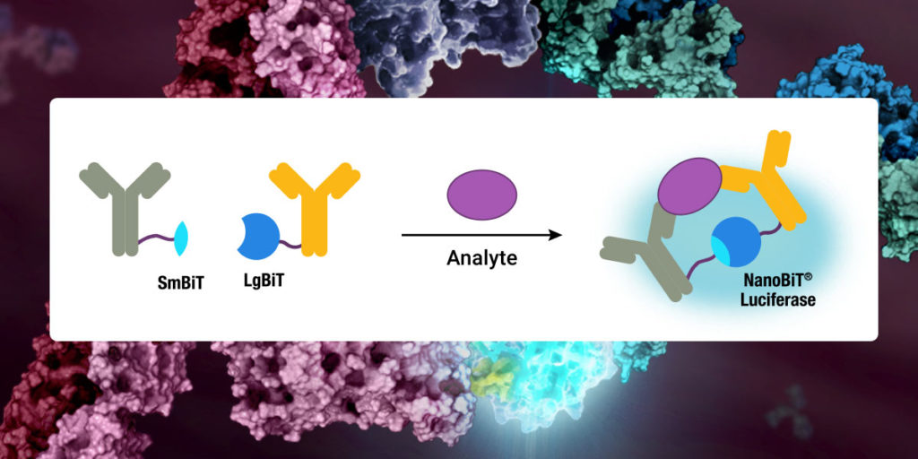
This diagram shows how the Lumit™ Immuno assay can be used to detect cytokines.

Cytokines—small signaling molecules that regulate inflammation and immunity—have recently become the focus of inflammation research due to their role in causing severe COVID-19 symptoms. In these severe cases, the patient’s immune system responds to the infection with uncontrolled cytokine release and immune cell activation, called the “cytokine storm”. Although the cytokine storm can be treated using established drugs, more research is needed to understand what causes this severe immune response and why only some patients develop it.

Continue reading “Understanding Inflammation: A Faster, Easier Way to Detect Cytokines in Cells”