Oncolytic Viruses: Models and Assays for Developing Viruses That Can Kill Cancer

When we think of viruses, we often think of diseases, pandemics and death. Our impression of viruses is that they are “bad”. But viruses could also be a possible cure for the deadliest disease in modern history: cancer. The therapeutic effects of “good” cancer-killing oncolytic viruses have been documented over a century ago. Records from as early as 1904 described a 42-year old woman with acute leukemia who experienced temporary remission after an influenza infection. Other early reports showed spontaneous remission of Hodgkin lymphoma and Burkitt’s lymphoma after natural infections with the measles virus.

Despite the long history, oncolytic viruses have only recently gained momentum in the scientific community. Dr. Aldo Pourchet, CSO and co-founder of Omios Biologics—a biotech startup in the San Francisco Bay area—is determined to harness the power of oncolytic viruses to develop a new generation of cancer immunotherapy.

How Oncolytic Viruses Work

“One thing that we know for sure is that you need the immune system to fight the cancer,” says Pourchet. “You need to recruit the immune system, and probably the best thing we know for recruiting the immune system is viruses. Our immune system evolved to detect them immediately. That’s why we are still on Earth. It’s because we have been able to fight deadly viruses.”

Continue reading “Oncolytic Viruses: Models and Assays for Developing Viruses That Can Kill Cancer”
Reaching Out for Lab Research Experience

Today’s guest blog is written by Melissa Martin, a global marketing intern with Promega this summer. She will be a senior this fall at the University of Wisconsin-Madison where she is double majoring in zoology and life sciences communication, with a certificate in environmental studies.

Congrats! You are attending a university and pursuing a challenging, yet rewarding, undergraduate science degree. Getting to this moment probably included lots of late nights spent studying or worrying while applying to your dream college. However, now that you are here you will find that classes provide a lot of information. You can even take your education one step further by getting hands-on experience in a research lab.

Working in a lab is not only about making your resume look good. It offers a real-world experience that directly enhances your learning experience and can even guide your future. For example, your experiences in the lab can teach you basic skills (pipetting, determining concentrations, performing titrations, etc.) that will be useful in a variety of science professions.

Continue reading “Reaching Out for Lab Research Experience”

Immune Checkpoint Bioassays Strengthen Cancer Research in the Development of New Therapies

This post was written by guest blogger, Nicole Werner, Product Management Support at Promega GmbH.

“You have cancer.” – a statement that fundamentally changes life in a second. After the first shock, the insight often arises: “If only I had stopped smoking sooner!”

Lung cancer, while not the leading cause of death worldwide, is the leading preventable cause of death in developed countries. According to the WHO, eight million people die each year as a result of smoking, including one million as a result of passive smoking [1]. Currently, 80% of those affected die within the next 13 months after diagnosis [1]. New therapeutic approaches, such as treatment with immune checkpoint inhibitors, bring hope.

Promega supports research in this area with the high-precision tools needed to develop this new form of therapy.

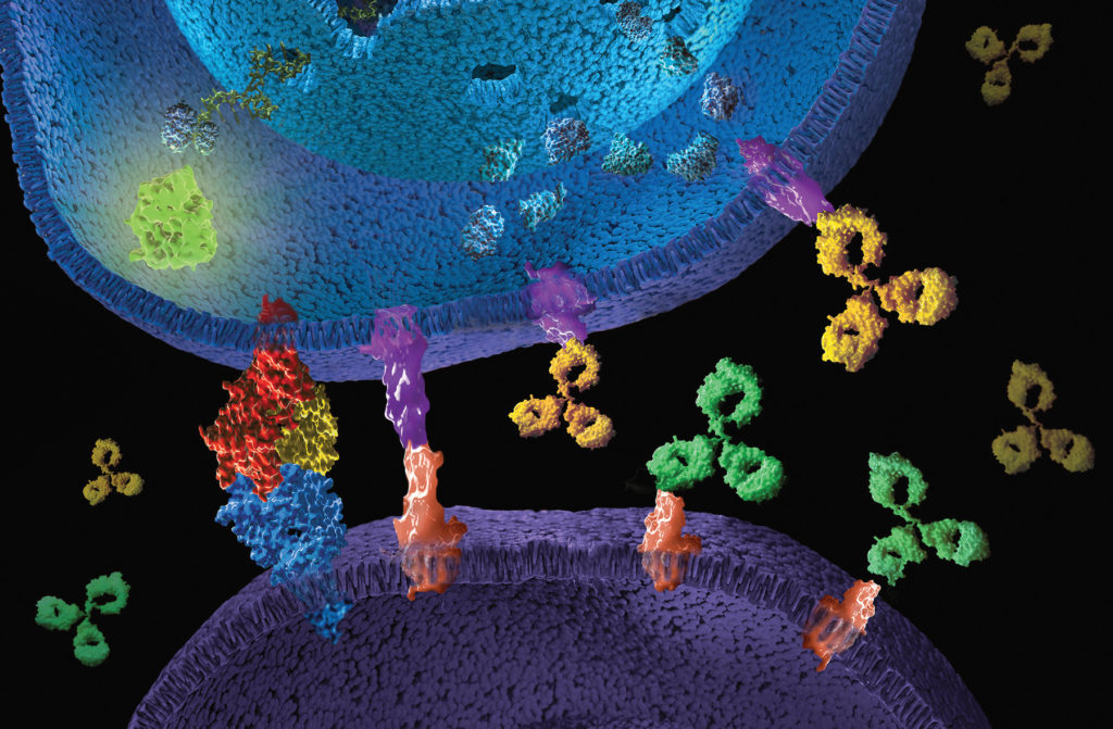
Artistic 3D rendering  of Immune checkpoint signaling. Immune checkpoint bioassays enable researcher to characterize  therapeutic antibodies trageting these pathways.
Continue reading “Immune Checkpoint Bioassays Strengthen Cancer Research in the Development of New Therapies”

RIPK1: Promising Drug Target of Chronic Inflammatory Diseases

Today’s post is written by Michael Curtin, Senior Product Manager, Reporters and Signaling.

Inflammation is a defense mechanism that the body employs in which the immune system recognizes and removes harmful and foreign stimuli and begins the healing process. Inflammation can be either acute or chronic. Chronic inflammation is also referred to as slow, long-term inflammation and can last for prolonged periods (several months to years); chronic inflammation is caused by immune dysregulation. This typically takes the form of the body’s inability to resolve inflammation resulting from overproduction of inflammatory cytokines and chemokines, as well as danger-associated molecular patterns (DAMPs) released from dying cells (2). Tumor Necrosis Factor (TNF) is the primary cytokine involved in many common inflammatory diseases and is where many therapies targeting inflammation are focused.

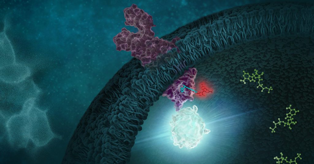
Signaling of kinases like RIPK1 can be studied using the NanoBRET target engagement assays

Recent research that RIP kinases (RIPK1 and RIPK3) are important regulators of innate immunity via their key roles in cell death signaling during cellular stress and following exposure to inflammatory and infectious stimuli. RIPK1 has an important scaffolding role in pro-inflammatory signaling where it interacts with TRADD, TRAF1 TRAF2, and TRAF3 and TRADD can act as an adaptor protein to recruit RIPK1 to the TNFR1 complex in a TNF-dependent process. RIPK1 plays a kinase activity-dependent role in both apoptotic and necroptotic cell death. A review article by Speir et al. (1) discusses the role of RIP kinases in chronic inflammation and the potential of RIPK1 inhibitors as a new therapeutic approach for the treatment of chronic inflammation. RIPK1 or Receptor Interacting Protein Kinase 1 is a serine/threonine kinase that was originally identified as interacting with the cytoplasmic domain of FAS. Promega offers several reagents that make studying RIPK1 easier- these include our RIPK1 Kinase Enzyme Systems which includes RIPK1 (Human, recombinant; amino acids 1-327), myelin basic protein (MBP) substrate, reaction buffer, MnCl2, and DTT and is optimized for use with our ADP-Glo Kinase Assay.

Continue reading “RIPK1: Promising Drug Target of Chronic Inflammatory Diseases”

Overcoming Challenges to Detect Apoptosis in 3D Cell Structures

This blog is written by guest author, Maggie Bach, Sr. Product Manager, Promega Corporation.

Researchers are increasingly relying on cells grown in three-dimensional (3D) structures to help answer their research questions. Monolayer, or 2D cell culture, was the go-to cell culture method for the past century. Now, the need to better represent in vivo conditions is driving the adoption of 3D cell culture models. Cells grown in 3D structures better mimic tissue-like structures, better exhibit differentiated cellular functions, and better predict in vivo responses to drug treatment.

Switching to 3D cell culture models comes with challenges. Methods to interrogate these models need to be adaptable and reliable for the many types of 3D models. Some of the most popular 3D models include spheroids grown in ultra-low attachment plates, and cells grown in an extracellular matrix, such as Matrigel® from Corning. Even more complex models include medium flow over the cells in microfluidic or organ-on-a-chip devices. Will an assay originally developed for cells grown in monolayer perform consistently with various 3D models? How is measuring a cellular marker different when cells are grown in 3D models compared to monolayer growth?

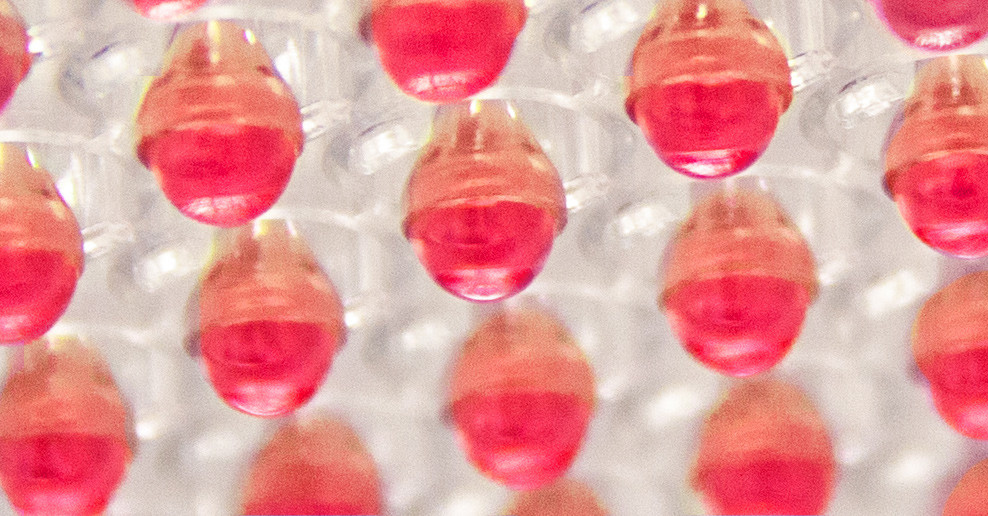
Close up of cells in 3D culture apparatus. 3D Cell Structures Provide Challenges for Measuring Markers of Cellular Activitiy
3D Cell Structures Provide Challenges for Measuring Markers of Cellular Activitiy
Continue reading “Overcoming Challenges to Detect Apoptosis in 3D Cell Structures”

World Firefly Day: Shining More Light on Glo-ing Innovations

On July 3rd and 4th, 2021, we celebrate World Firefly Day, and 2021, marks 30 years of luciferase products firefly luciferase vectors and Luciferase Assay System. These tools are key in advancing bioluminescent technology. To celebrate this day, we want to highlight some innovations that have been made possible with these tools.

Continue reading “World Firefly Day: Shining More Light on Glo-ing Innovations”

Diversifying Biotech: D.O.O.R.S. Scholarship Empowers Young Scientists

The DOORS Scholarship stands for Diversification of our Research Scientists.

In 2020, Promega North America launched the Diversification Of Our Research Scientists (DOORS) Scholarship to recognize and empower students from underrepresented backgrounds. Ten students received $5,000 towards tuition and other costs associated with their education, as well as connections with mentors from Promega. Here are two of their stories.

Continue reading “Diversifying Biotech: D.O.O.R.S. Scholarship Empowers Young Scientists”

RAS-Targeted Drug Discovery: From Challenge to Opportunity

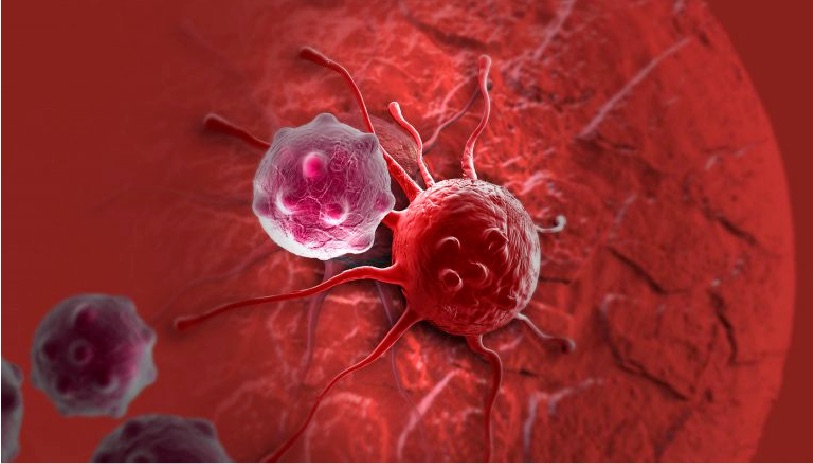
cancer cell, ras-targeted drug discovery

In 1963, Jennifer Harvey was studying Moloney murine leukemia virus (MMLV) at the cancer research department of the London Hospital Research Laboratories. After routine transfers of plasma from MMLV-infected rats to mice, she made an unusual discovery. In addition to the expected leukemia, the mice that received the plasma developed solid tumors (soft-tissue sarcomas), primarily in the spleen (1). A few years later, Werner Kirsten at the University of Chicago observed similar results working with mouse erythroblastosis virus (MEV) (2).

Subsequent research, with the advent of genome sequencing, showed that a cellular rat gene had been incorporated into the viral genome in both cases (3). These genomic sequences contained a mutation later shown to be responsible for the development of sarcomas, and the word “oncogene” became a common part of the vocabulary in cancer publications during the early 1980s (4). Harvey’s discovery led to the naming of the corresponding rat sarcoma oncogene as HRAS, while Kirsten’s related oncogene was named KRAS. Several laboratories, working independently, cloned the human homolog of the viral HRAS gene in 1982 (3). The human KRAS gene was cloned shortly thereafter, as well as a third RAS gene, named NRAS (3). Additional studies showed that a single point mutation in each of these genes led to oncogenic activation, and they have been popular targets for anticancer drug discovery efforts ever since.

Continue reading “RAS-Targeted Drug Discovery: From Challenge to Opportunity”

Antibody Response Differ in Adults, Kids and Potential Cross-Reactive Coronavirus Antibodies

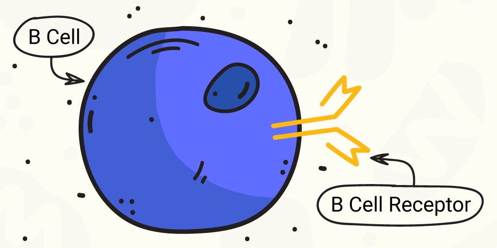
B cell and B cell receptor cartoon. The B cell receptors are important for antibody response.
Drawing of a B cell and the B cell receptor. The receptor shows the characteristic Y shape of an immunoglobulin molecule.

B cells are the immune cells that produce antibodies (immunoglobulins or Ig) to detect intruding pathogens. B cells produce a variety of classes of antibodies. Generally during an immune response to a pathogen, whether viral or bacterial, B cells produce immunoglobulins (Ig) IgM and IgD, and later in the response, IgG and IgA, that are specific to the intruding organism. These Igs capture and aid in neutralizing the pathogen.

Ig classes can be studied by sequencing the B cell receptor (BCR), which binds antigen specifically. BCRs are formed via irreversible gene segment rearrangements of variable, diversity and joining (VDJ) genes. Ig classes can be diversified through somatic hypermutation and class-switch recombination of these gene segments (1).

B cell receptors with high sequence similarity can be found in individuals exposed to the same antigen, demonstrating that antigen exposure can result in similar B cell clones and memory B cells between individuals, both adults and children (1).

However, B cell immune responses can differ between adults and children. For example, children use more B cell clones that form neutralizing antibodies to HIV-1. And children infected with SARS-CoV-2 generally have milder illness than infected adults. SARS-CoV-2-infected children have lower antibody titers to the virus and more IgG-specific response to SARS-CoV-2 spike protein than to the nucleocapsid protein (1). These differences can contribute to faster SARS-CoV-2 clearance and lower viral loads in children versus adults.

Continue reading “Antibody Response Differ in Adults, Kids and Potential Cross-Reactive Coronavirus Antibodies”

Questions Arise about TMB as a Predictive Biomarker for Immune Checkpoint Inhibitor Therapy

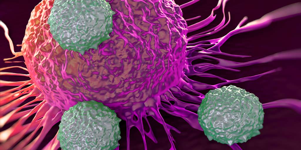
Artist rendition of immune cells attacking a cancer cells. Immune checkpoint inhbitor therapy is a relatively new therapy for certain cancers.

Immune checkpoint inhibitor (ICI), or immune checkpoint blockade, therapies are a revolutionary, and relatively new, approach to treating cancer. These therapies work by blocking immune checkpoint proteins that act to negatively regulate the immune system through the PD-1 pathway. Some tumors express immune checkpoints to prevent the immune system from producing a strong enough immune response to kill the cancer cells. When these checkpoint proteins are blocked by an ICI, the body’s T-cells can recognize and kill the cancer cells. ICI therapies show tremendous promise. Unfortunately, not all tumors express immune checkpoint proteins, and so, not all tumors will be effectively treated with ICI therapies. The challenge is differentiating between the tumors that will respond and tumors that won’t.

DNA Mismatch Repair Deficiency Status as Detected by Microsatellite Instability or Immunohisotchemistry are Important Biomarkers for ICI

Biomarkers are measurable indicators of a clinical condition that can be found in tissue, blood, or other fluids. Predictive biomarkers for ICIs can help determine if these therapies are a suitable choice for treatment. Some tumors have deficiencies in their DNA mismatch repair mechanisms. Mismatch repair deficiency (dMMR) leads to the accumulation of mutations across the genome, particularly in microsatellites, which over time can result in higher levels of neoantigen production, rendering the tumors susceptible to the ICI therapy (1–5).

In 2017, Le et al. demonstrated that dMMR status reliably predicted response to an ICI therapy targeting the PD-1 checkpoint protein (6). Following this discovery, ICI based on dMMR  determined using either microsatellite instbility (MSI) or immunohistochemistry (IHC), gained clearance from the US Food and Drug Administation (FDA) for microsatellite instability-high (MSI-H) or dMMR by IHC solid tumors. This was the first time a cancer treatment was cleared based on a biomarker regardless of cancer origin (1,7).  Since then, MSI-H and dMMR, have become some of the most recognized tissue agnostic biomarkers for improved survival following ICI therapy of solid tumors (6,8,9).

Continue reading “Questions Arise about TMB as a Predictive Biomarker for Immune Checkpoint Inhibitor Therapy”