Evidence of Inflammasome Activation in Severe COVID-19

The pandemic caused by SARS-CoV-2 has brought the world to its knees. There have been many deaths, many persons with lingering disease (long COVID) and the inability to vaccinate everyone quickly, for starters. SARS-CoV-2 has not only been a tricky adversary in terms of treatment options to save lives, it’s also been a wily opponent to researchers studying the virus.
Contributing to the existing studies, with their review of the role of inflammasomes in COVID-19, Vora et al. recently published “Inflammasome activation at the crux of severe COVID-19” in Nature Reviews Immunology. In this paper they detail evidence of inflammasome activation and its role in SARS-CoV-2 infections.

Contributions of Those Lost in the SARS-CoV-2 Pandemic
I’d like to take a moment to note the uniquely awful nature of the virus at the center of this blog and the paper it reviews. Many of the papers we blog about describe research involving cell lines, mice or another animal model. The closest most reports get to human research subjects is the use of human cells lines. In the Vora et al. report, serum and tissue samples are from actual human patients, some that survived and many that did not survive COVID-19. It’s not lost on us, Dear Reader, the contributions of those that suffered and died due to SARS-CoV-2 infection. Many persons with severe or fatal COVID-19 have made a significant contribution to our understanding of this virus and its treatment options. We owe them, as well as the researchers that have studied SARS-CoV-2, our sincerest gratitude.

Why the Interest in Inflammasomes?
For detailed information on inflammasomes you can read Ken’s blog, here. You will find background information there and on our inflammasome web page.

In their paper, Vora et al. provide evidence of inflammasome activation, both direct and indirect, in COVID-19. The authors note:

“Key to inflammation and innate immunity, inflammasomes are large, micrometrescale multiprotein cytosolic complexes that assemble in response to pathogen-associated molecular patterns (PAMPs) or damage-associated molecular patterns (DAMPs) and trigger proinflammatory cytokine release as well as pyroptosis, a proinflammatory lytic cell death.”

Continue reading “Evidence of Inflammasome Activation in Severe COVID-19”

New Therapy for Brain Tumors: High-Pressure Oxygen Rewires Glucose Metabolism in Glioblastoma

Glioblastoma (GBM) is an aggressive type of brain tumor, and one of the deadliest cancers. GBM is often treated with surgery, radiation and chemotherapy, but even if the initial treatment is successful, a majority of patients relapse within months. One reason why GBM is so difficult to treat is the hypoxic (low-oxygen) tumor environment. It is known that hypoxic cells are resistant to radiotherapy; the greater the number of tumor stem cells in a hypoxic environment, the less efficient radiotherapy is at controlling tumor growth.

A new therapeutic approach aims to remove the hypoxic environment in GBM by administering pure oxygen to patients at high pressure, known as “hyperbaric oxygen (HBO) therapy”. Previous studies have shown that HBO improves the efficacy of radiotherapy in GBM patients. However, the therapeutic mechanism of HBO was largely unknown. That is, until now.

Dr. Anna Tesei, the Head of Radiobiomics and Drug Discovery at the Biosciences Laboratory of IRST-IRCCS in Italy, recently published a study on the mechanism in which HBOT affects GBM tumor cells and the tumor environment. “The main purpose of our study was to provide a preclinical rationale for the use of hyperbaric oxygen in association with radiotherapy for the treatment of GBM,” she says.

Dr. Anna Tesei is studing glucose metabolism in glioblastoma cells.
Continue reading “New Therapy for Brain Tumors: High-Pressure Oxygen Rewires Glucose Metabolism in Glioblastoma”

Seeing is Believing: How NanoLuc® Luciferase Illuminates Virus Infections

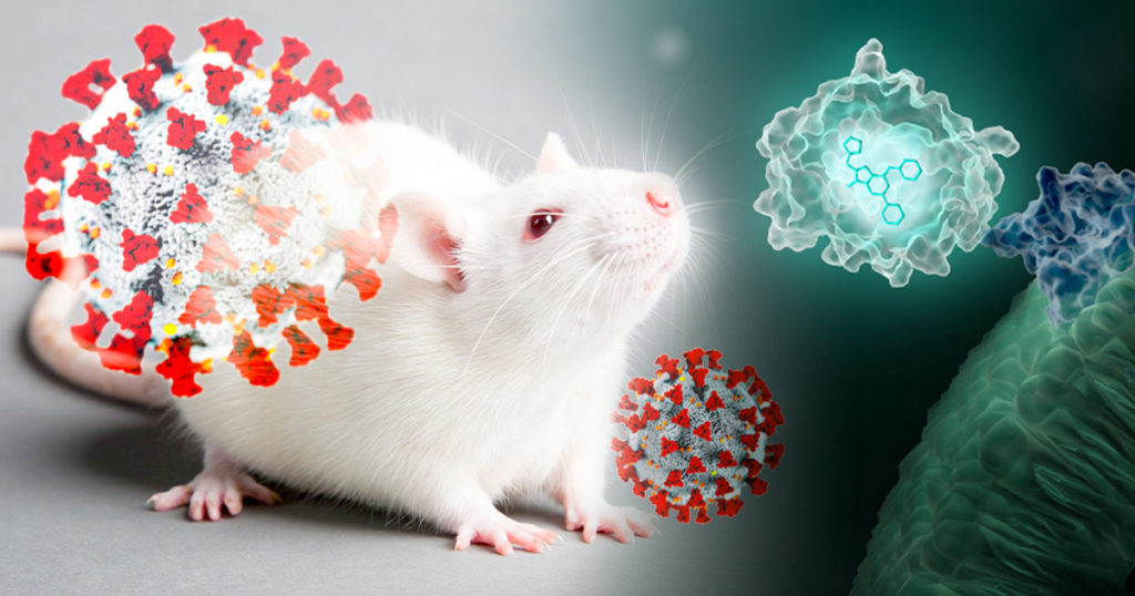
Artists interpretation of in vivo imaging of viral infections in mice using NanoLuc luciferase.

Wearing blue surgical gowns and white respirator hoods, research scientist Pradeep Uchil and post-doctoral fellow Irfan Ullah carry an anesthetized mouse to the lab’s imaging unit. Two days ago, the mouse was infected with a SARS-CoV-2 virus engineered to produce a bioluminescent protein. After an injection of a bioluminescence substrate, a blue glow starts to emanate from within the mouse’s nasal cavity and chest, visible to the imaging unit’s camera and Uchil’s eyes.

“We were never able to see this kind of signal with retrovirus infections.” Uchil is a research scientist at the Yale School of Medicine whose work focuses on the in vivo imaging of retroviral infections. Normally, the mouse would have to be sacrificed and “opened up” for viral bioluminescent signals from internal tissues to be imaged directly.

Continue reading “Seeing is Believing: How NanoLuc® Luciferase Illuminates Virus Infections”

Automating Forensic DNA Purification to Meet Urgent Needs: Reflections on September 11, 2001

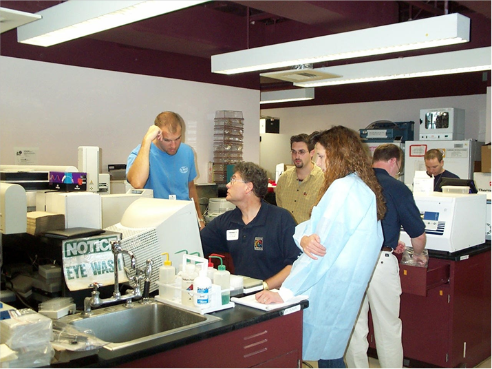
Allan Tereba (center, blue polo) works with technicians at the New York City Office of the Chief Medical Examiner (OCME) in September 2001 to discuss automating forensic DNA purificaiton.
Allan Tereba (center, blue polo) works with technicians at the New York City Office of Chief Medical Examiner (OCME) in September 2001.

In the summer of 2000, Promega research scientist Allan Tereba was asked to develop an automated protocol for purifying DNA for forensics. His team had recently launched DNA IQ, the first Promega kit for purifying forensic DNA using magnetic beads. This was before the Maxwell® instruments, and before Promega purification chemistries were widely adaptable to high-throughput automation.

“I had my doubts about being able to do that,” Allan says. “When you’re working with STRs, small amounts of contaminant DNA are going to mess up your results. But I went ahead and tried it, and it was a challenge.”

A little over a year later, Allan was in his office when he heard on the radio that a plane had struck the North tower of the World Trade Center in New York City. Shortly after, he heard the announcement that a second plane had hit the South tower.

By that point, Allan and his colleagues had successfully adapted DNA IQ to be used on the deck of a robot. Within days of the attacks, Promega scientists were supporting the New York City Office of Chief Medical Examiner (OCME) and New York State Police in their work to identify human remains that were recovered from Ground Zero.

Thanks to the work of Allan and many other Promega scientists, Promega was prepared to offer unique solutions to urgent needs. In their own words, here are some of those scientists’ reflections.

Continue reading “Automating Forensic DNA Purification to Meet Urgent Needs: Reflections on September 11, 2001”

The Stories in the Bones: DNA Forensic Analysis 20 Years after 9/11

September 11, 2001 is the day that will live in infamy for my generation. On that beautiful late summer day, I was at my desk working on the Fall issue of Neural Notes magazine when a colleague learned of the first plane hitting the World Trade Center. As the morning wore on, we learned quickly that it wasn’t just one plane, and it wasn’t just the World Trade Center.

Two beams of light recognized the site of the World Trade Center attack. Today DNA forensic analysis applies new technologies to bring closure to families of victims.

Information was sparse. The world wide web was incredibly slow, and social media wasn’t much of a thing—nothing more than a few listservs for the life sciences. Someone managed to find a TV with a rabbit-eared, foil-covered antenna, and we gathered in the cafeteria of Promega headquarters—our shock growing as more footage became available. At Promega, conversation immediately turned to how we could bring our DNA forensic analysis expertise to help and support the authorities with the identification of victims and cataloguing of reference samples.

Just as the internet and social media have evolved into faster and more powerful means of communication—no longer do we rely on TVs with antennas for breaking news—the technology that is used to identify victims of a tragedy from partial remains like bone fragments and teeth has also evolved to be faster and more powerful.

Teeth and Bones: Then and Now

“Bones tell me the story of a person’s life—how old they were, what their gender was, their ancestral background.”  Kathy Reichs

Many stories, both fact and fiction, start with a discovery of bones from a burial site or other scene. Bones can be recovered from harsh environments, having been exposed to extreme heat, time, acidic soils, swamps, chemicals, animal activities, water, or fires and explosions. These exposures degrade the sample and make recovering DNA from the cells deep within the bone matrix difficult.

Continue reading “The Stories in the Bones: DNA Forensic Analysis 20 Years after 9/11”

New Cleared IVD Assay for Microsatellite Instability in Colorectal Cancer Aims to Help Identify Those with Lynch Syndrome

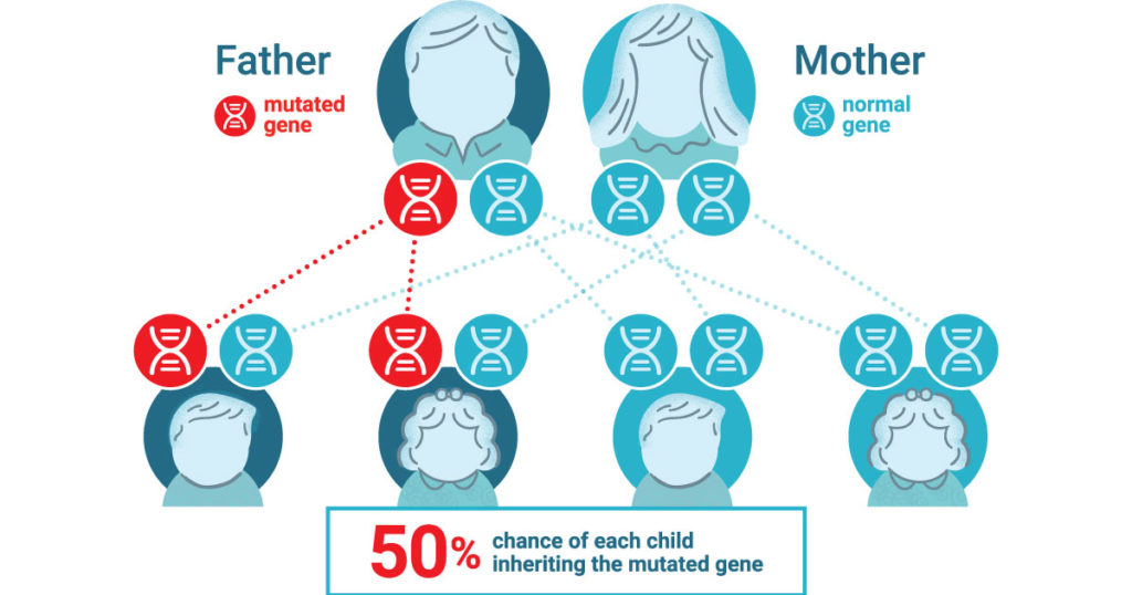
Lynch syndrome is an inherited condition that significantly increases the risk of developing colorectal and other cancers, often at a young age. People with this condition have close to an 80% chance of developing colorectal cancer in their lifetime. It is the most common form of hereditary colon cancer and causes roughly 3% of all colon cancers. The mutations that cause Lynch syndrome are inherited in an autosomal dominant manner— ­meaning you only need to have one copy of the gene with a Lynch-associated mutation to be at an increased risk.

It is estimated that 1 in every 279 people have inherited a Lynch-associated mutation (1). Yet despite this prevelence, Lynch syndrome is not well known and ~95% of those with the syndrome don’t know they have it (1).

Lynch Syndrome Cause and Detection

Lynch syndrome is caused by mutations that result in the loss of function of one of the four different major mismatch repair proteins. These proteins act as “proof readers” that correct errors in the DNA sequence that can occur during DNA replication. To determine if Lynch syndrome is likely, simple screening tests can be performed on tumor (cancer) tissue to indicate if more specific genetic testing should be considered. One such screening looks for high levels of microsatellite instability (MSI) in the tumor tissue. High microsatellite instability (MSI-H) in tumor tissue is a functional indication that one or more of the major mismatch repair proteins is not functioning properly.

Watch this short video to learn more about microsatellite instability.

For those who develop colorectal cancer at an early age or have a family history (immediate family member or multiple family members with colorectal cancer or polyps), screening for Lynch syndrome can offer valuable insight for both patients and their family, as well as for their healthcare provider.

New MSI IVD Test for Colorectal Cancer to Help Identify Lynch Syndrome

The newly released Promega OncoMate® MSI Dx Analysis System is an FDA-cleared IVD Medical Device and can be used to determine the MSI status of colorectal cancer tumors to aid in identifying those who should be further tested for Lynch syndrome. The OncoMate MSI Dx Analysis System builds upon the company’s fifteen year history of supporting global cancer researchers with one of the leading standard tests for MSI status detection. The OncoMate MSI Dx Analysis System offers an improved formulation while using the same five markers that have become the gold standard for MSI detection in the research community and is referenced in over 140 peer review publications (2,3).

The OncoMate® MSI Dx Analysis System is designed to provide physicians with a functional, molecular measurement of the level of DNA mismatch repair deficiency demonstrated within their patient’s colorectal cancer tumor. MSI testing is recommended to identify candidates for further diagnostic testing for Lynch syndrome. (2–4). The System is part of a broader workflow that includes DNA extraction from FFPE tissue samples, quantitation of DNA, amplification of specific microsatellite markers using multiplex PCR, fragment separation by capillary electrophoresis, and data analysis and interpretation software. The OncoMate MSI Dx Analysis System is available in certain countries.  Visit the OncoMate MSI Dx Analysis System webpage to learn more.

Promega previously announced a CE-marked version of the OncoMate MSI  Dx Analysis System in France, Germany, Austria, Poland, UK, Ireland, Belgium, Netherlands, Luxembourg, Spain, Italy, Switzerland, Denmark, Sweden and Norway.

For more information about MSI solutions available from Promega visit our Microsatellite Instability Testing webpage.

References

  1. Win, A. K. et al. (2017) Cancer Epidemiol. Prev. 26, 404–12.
  2. Bacher, J. et al. (2004) Dis. Markers 20, 237–50.
  3. Svrek, M. et al. (2019) Bull. Cancer, 106, 119–28.
  4. Umar, A. et al. (2004) J. Natl. Cancer Inst. 18, 261–8.

National Wildlife Day: Admiring Our Natural World

On September 4th, 2021 we celebrate National Wildlife Day. This day helps cherish our planet’s biodiversity and recognize issues that impact wildlife. Take a look at three Promega blogs that highlight preservation and conservation efforts being made to support our natural world.

Continue reading “National Wildlife Day: Admiring Our Natural World”

Mentally Preparing for My Return to the Lab: A Grad Student’s Perspective

Today’s guest blog is written by Sophie Mancha, a global marketing intern with Promega this summer. She will be starting her 4th year as a PhD candidate in the Biomedical Engineering Department at the University of Wisconsin-Madison, studying pancreatic cancer.

Graduate students are used to working. Not only during regular work hours but also well into the night to finish readings or work on data analysis. Ripping graduate students away from their research as they desperately try to produce useful data may be as hard as finding toilet paper during the first few months of the SARS-CoV-2 outbreak. However, across the world graduate students saw their research come to a screeching halt. The pandemic took over and everyone suddenly went into quarantine.

I clearly remember my first virtual lab meeting. We all frantically tried figuring out what video-conferencing platform to use and how to share our screens. We kept repeating “stay calm” as we naively thought this would only last a couple of weeks. As the months went by, I began to panic. I realized I had finished analyzing the last remaining data I had left and was no longer being “productive”. This quickly spiraled into thoughts that I may never earn my PhD.

Continue reading “Mentally Preparing for My Return to the Lab: A Grad Student’s Perspective”

LncRNA: The Long and Short of “Junk RNA”

The Central Dogma and Junk DNA

lncRNA, long noncoding RNA

On September 19, 1957, Francis Crick delivered a lecture during a symposium at University College London, titled “Protein Synthesis”. The lecture was published a year later (1); in it, Crick quotes his colleague James Watson as saying, “The most significant thing about the nucleic acids is that we don’t know what they can do.” In contrast, Crick argued that proteins play a central, indispensable role as enzymes within the cell that catalyze a variety of chemical reactions. He believed that the main role of genetic material was to control the synthesis of proteins, although the mechanism of that process was not known.

Crick’s hypothesis came to be known as the central dogma of molecular biology, and it was immortalized in his hand-written notes that described the flow of information from DNA to RNA to proteins. This achievement was all the more remarkable, considering that messenger RNAs were completely unknown at that time, and very little was known about how the cellular translational machinery functioned within the cytoplasm to synthesize proteins. Although the later discovery of retroviruses appeared to challenge Crick’s central dogma, he explained quite succinctly that his original statement had simply been misunderstood, and that information could flow in both directions between DNA and RNA (2).

Continue reading “LncRNA: The Long and Short of “Junk RNA””

Psilocybin as Antidepressant: Quick Acting, Lasting Benefits

Depression is not simply a mood disorder, a feeling of sadness, or being ill at ease. Depression can completely shut a person down, manifesting as an inability to make decisions, to take action, to think. Even sleep is affected by depression.

Researchers and clinicians who treat depression are learning that the physical manifestations can be mirrored by internal, cellular changes. Some people with depression have decreases in their gray matter volume, particularly in areas like the hippocampus (important to memory, learning, and emotions) and prefrontal cortex (where higher-level thought and planning abilities are based).

Additionally, imaging has shown a decrease in the number of synapses—the structures through which electrical or chemical signals are passed between neurons and other cells—in persons with chronic depression. Without the signals that synapses transmit, brain function is disrupted.

And without intervention in depression, synapse decrease can continue.

While there are drugs and behavioral therapies to treat depression, these therapies can be slow to act and sometimes ineffective. In addition, once synaptic loss has occurred, these therapies are less effective.

In their August 2021 paper, “Psilocybin induces rapid and persistent growth of dendritic spines in frontal cortex in vivo” (1), Shao et al. state,

“It has long been recognized that these compounds (serotonergic psychedelics like psilocybin) may have therapeutic potential for neuropsychiatric disorders, including depression, obsessive-compulsive disorder and addiction”.

Continue reading “Psilocybin as Antidepressant: Quick Acting, Lasting Benefits”