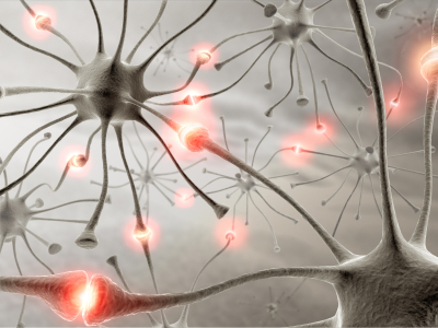
The brain is constantly rewiring itself, fine-tuning connections that shape how we think, learn, and remember. But capturing those fleeting molecular changes as they happen — at the level of individual synapses and across entire brain regions — has long been a challenge in neuroscience. Now, thanks to recent advances in HaloTag® dye technology, researchers can visualize protein dynamics in living brains with stunning clarity and specificity.
The challenge of Imaging Neuronal Processes
Understanding how proteins like AMPA receptors (a major class of excitatory glutamate receptors in the brain), and PSD-95 (a scaffolding protein found at excitatory synapses) move, accumulate, and degrade within neurons is essential to decoding the mechanisms of memory and learning. But many traditional imaging methods fall short when it comes to:
- Resolving synaptic events deep in tissue
- Monitoring protein turnover over time
- Linking molecular changes to behavioral outcomes
This is where HaloTag®-based pulse-chase labeling using Janelia Fluor® ligands shines. By combining genetic tagging with highly bioavailable, spectrally distinct fluorescent dyes, researchers can watch proteins come and go across the brain — in real time, in vivo, and at synaptic resolution. Here we highlight several recent publications that demonstrate novel applications using the powerful Janelia Fluor® dyes.
EPSILON: Watching Synapses Form Memories
In a recent study published in Nature Neuroscience, researchers developed a technique called Extracellular Protein Surface Labeling in Neurons (EPSILON) — a method for tracking AMPAR exocytosis, insertion of receptors into the cell membrane, in live mice1. AMPAR levels at synapses influence how strongly neurons communicate — a key process that changes during learning and memory formation, often referred to as long-term potentiation (LTP). Traditional methods for monitoring AMPAR dynamics have proven challenging, with no technique able to identify brain location, window of time, and specific neurons simultaneously. To track newly inserted receptors, the authors fused GluA1 to HaloTag® and performed sequential labeling with two impermeant dyes — first tagging receptors already on the cell surface, then labeling newly exposed receptors after AMPAR exocytosis. They used distinct colors to generate “snapshots” of synaptic potentiation during memory formations.
Building upon this, the authors explored how memories are formed by using a behavioral model called contextual fear conditioning, in which mice learn to associate a specific environment with a mild foot shock. When placed back in that environment later, mice display a conditioned freezing response — a common measure of learned fear. To track the activation of hippocampal CA1 cells which are involved in memory formation, they tracked the expression of an early gene used as a marker of neuronal activity, FOS. In this model, the EPSILON technique revealed:
- AMPAR exocytosis tracks memory-relevant neuronal activity, with a strong correlation between cFos expression and strengthening of synapse-level connections, or potentiation, in CA1 neurons during memory formation.
- Synaptic plasticity isn’t evenly distributed; it is spatially patterned and favors near the cell body and base of neurons (perisomatic and basal synapses), rather than at the far ends of dendritic branches (distal and apical spines).
- Potentiated spines, or stronger synapses, cluster in specific parts of a neuron, creating ‘hotspots’ of plasticity, and ‘silent’ dendritic segments. This suggests that memory encoding may be spatially concentrated within select dendritic branches, rather than uniformly distributed.
This method not only visualized learning-related plasticity with single-synapse precision but also provided a molecular link between immediate early gene expression and actual synaptic changes.
DELTA: Mapping Protein Turnover Brain-Wide
While EPSILON zooms in, DELTA zooms out — A second paper published in Nature Neuroscience offers a brain-wide view of protein turnover. Using HaloTag® knock-in mice and spectrally distinct Janelia Fluor® HaloTag® ligands, the authors utilized pulse-chase labeling of synaptic proteins to develop DELTA (Dye Estimation of the Lifetime of proTeins in the brAin) to capture how long specific synaptic proteins persist before being replaced2.
Pulse-chase labeling is a method that uses sequential dye labeling to track old vs. new proteins. The protein population labeled with the first dye ligand infusion is the “pulse”, and the newly synthesized or trafficked protein labeled with the second dye ligand infusion is the “chase”. The authors wanted to map learning-related synaptic plasticity using various behavioral tasks and environmental enrichment. Using DELTA to study protein dynamics in the hippocampus, the authors focused on several targets. These included PSD-95, a key scaffolding protein that helps organize receptors and signaling molecules at excitatory synapses, and GluA2, an ionotropic glutamate receptor modified during synaptic plasticity. Using the DELTA technique revealed:
- Learning induces highly localized synaptic protein turnover, with increased GluA2 dynamics specifically in hippocampal CA1, pointing to region- and task-specific plasticity during memory formation.
- Environmental enrichment triggers widespread synaptic remodeling, accelerating turnover of key synaptic proteins like PSD-95 and GluA2 across multiple brain regions, especially the neocortex.
- Synaptic protein turnover is spatially compartmentalized within neurons, with distinct turnover patterns in dendritic layers receiving different inputs, revealing input-specific regulation of plasticity at the subcellular level.
The DELTA technique enables researchers to match behavioral experiences with the molecular timeline of plasticity, helping bridge the gap between behavior, brain circuits, and modifications at the protein level.
HaloTag® Ligands: Powering a New Era of Brain Imaging
Both EPSILON and DELTA rely on the HaloTag® system combined with advanced Janelia Fluor® dyes — technologies supported by Promega. Our portfolio of membrane-impermeant and -permeant ligands allow researchers to label complex neuronal systems down to the synaptic level. These ligands enable the following:
- Label proteins both cellularly and in vivo with high specificity and a high signal-to-noise ratio for bright signal with minimal background
- Label a wide variety of targets with cell-permeant and cell-impermeant ligands
- Correlate protein turnover with behavior, gene expression, or disease
- Compatible with advanced imaging techniques, such as pulse-chase experiments and super resolution microscopy
With rising demand for multiplexed, long-lasting, and cost-effective imaging tools in neuroscience, Promega’s HaloTag® imaging solutions are uniquely positioned to support cutting-edge research — from cell culture, to organotypic models, to live animal studies.
Request access to our in vivo HaloTag Ligands, available through early access.
Imaging the Future of Neuroscience
From revealing how memories take shape to tracking protein dynamics across the brain, in vivo imaging with HaloTag® dyes is unlocking new frontiers in neuroscience. As the field continues to explore ever more biologically relevant systems, HaloTag® Ligands and Janelia Fluor® dyes will remain indispensable tools for visualizing the complexity of the brain — one synapse at a time.
Citations:
Kim, D., et al. (2025). EPSILON: a method for pulse-chase labeling to probe synaptic AMPAR exocytosis during memory formation. Nature Neuroscience. https://doi.org/10.1038/s41593-025-01922-5
Mohar, B., et al. (2025). DELTA: a method for brain-wide measurement of synaptic protein turnover reveals localized plasticity during learning. Nature Neuroscience. https://doi.org/10.1038/s41593-025-01923-4