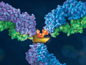
Therapeutic monoclonal antibodies (MAbs) are inherently heterogeneous due to a wide range of both enzymatic and chemical modifications, such as oxidation, deamidation and glycosylation which may occur during expression, purification or storage. For identification and functional evaluation of these modifications, stability studies
are typically performed by employing stress conditions such as exposure to chemical oxidizers, elevated pH and temperature.
To characterize MAbs, a variety of analytical techniques are chosen, such as size exclusion chromatography and ion exchange chromatography. However, due to the large size of the intact MAbs, these methods lack structural resolution. Often, the chromatographic peaks resolved by SEC and IEC methods are collected and further analyzed by peptide mapping to obtain more detailed information. Peptide mapping, in which antibodies are cleaved into small peptides through protease digestion followed by LC–MS/MS analysis, is generally the method of choice for detection and quantitation of site-specific modifications. However sample preparation and lengthy chromatographic separation make peptide mapping impractical for the analysis of large numbers of samples. In contrast to peptide mapping analysis, the middle-down approach offers the advantage of high-throughput and specificity for antibody characterization.
Limited proteolysis of IgG molecules by the IdeS enzyme has been introduced for antibody characterization due to its high cleavage specificity and simple digestion procedure.
IdeS cleaves the IgG heavy chain below the hinge region constant sequence ELLGGPS. After disulfide bonds reduction, this cleavage leads to the release of the three fragments of about 25 kDa is size. Analyses of IdeS digested MAbs by RPLC–MS has been reported for the identification of domain-specific oxidation, charge profiling and N-glycan profiling (1). In this study, separation of the domains and related variants was achieved in a 30-minute gradient. Domain specific oxidations were quantified by UV peak areas, no orthogonal methods were evaluated for comparison.
Are you looking for proteases to use in your research?
Explore our portfolio of proteases today.
In a recent publication (2) describes the development of a rapid and improved analytical method employing IdeS digestion, RP-UHPLC separation, UV and MS detection for antibody characterization. IdeS digestion conditions and UHPLC parameters used to separate domain-specific polypeptides generated by IdeS digestion,was systematically evaluated. Separation of the domains and related variants was achieved in 10 min using UHPLC reversed-phase column. Oxidation levels were compared with the values obtained by the more complicated and time-consuming peptide mapping method. The similar trends observed by the two methods demonstrated that the IdeS-UHPLC method is valuable as a higher throughput alternative to peptide mapping for monitoring modifications.
- An, Y., et al. (2014) A new tool for monoclonal antibody analysis-application of IdeS proteolysis in IgG domain-specific characterization. MAbs 6, 879–893.
- Zhang, B. et al. (2016) Development of a rapid RP-UHPLC–MS method for analysis of modifications in therapeutic monoclonal antibodies. Journal of Chromatography B, 1032, 172-81.
