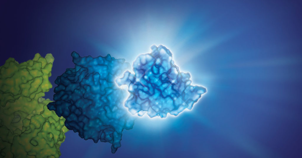
Bioluminescence imaging is a powerful tool for non-invasive studies of the effect of treatments on cells and tissues. The luminescent signal is strong, and can be used in vivo, enabling repeated observations over time, allowing longitudinal study of cellular changes for hours or days. Bioluminescence imaging can be used in live animals over varying periods of time, without interfering with normal cellular processes.
Fluorescence imaging is also used in cellular studies. Although it can provide a stronger signal than luminescence, fluorescence requires light for excitation, and thus its in vivo use is limited at a tissue or cell depth greater than 1mm.

In addition, autofluorescence can be an issue with fluorescence imaging, as cellular components and surrounding proteins and cells can fluoresce when exposed to light. Autofluorescence can result in high background signals, making it difficult to distinguish true fluorescence from background.
Because luminescence requires the enzyme luciferase and its substrate, luciferin, luminescence can have negligible background signaling. This high signal:low background ratio results in enhanced sensitivity compared to fluorescence imaging.
For two decades bioluminescence has been used for imaging in small mammal disease models. But attempts to use bioluminescence for imaging of brain tissues, has bumped up against the blood-brain barrier roadblock.
Luciferase and the Blood-Brain Barrier
D-luciferin, the substrate for firefly luciferase, is a small molecule that was previously thought to be blood-brain barrier permeable. However, recent studies with intraperitoneal administration of D-luciferin showed a very low signal from brain and central nervous system tissues (1).
The marine-based luciferase, NanoLuc® Luciferase, provides an alternative to firefly luciferase for in vivo studies and seen increased use in various areas of biomedical research, including in vivo tracking of viruses, exosomes and engineered CAR T-cells. Furimazine is the substrate for NanoLuc® Luciferase and the furimazine analog fluorofurimazine has been specifically optimized for in vivo measurement of NanoLuc® activity. Fluorofurimazine demonstrates maximal brightness in vivo, as well as good solubility and bioavailability. Read more about in vivo applications of NanoLuc® technology and the development of fluorofurimazine in the blog NanoLuc® Luciferase: Brighter Days Ahead for In Vivo Imaging.
To increase the utility of NanoLuc® as an in vivo reporter, steps were taken to quantify and optimize the abilities of furimazine for brain bioluminescent imaging.
NanoLuc® Substrate and the Blood-Brain Barrier
Su et al., in a recent publication in Nature Chemical Biology, report the results of evaluating and optimizing NanoLuc® substrates in brain bioluminescent imaging (1). They used mice genetically altered to express NanoLuc® Luciferase fused to fluorescent tags (Antares) in brain tissue, which enabled screening and characterizing of substrates furimazine, hydrofurimazine and fluorofurimazine, as well as structural analogs of these NanoLuc® substrates.
Their work showed that fluorofurimazine and hydrofurimazine performed less well in brain imaging than furimazine. This led the authors to tweak furimazine, including removal of hydroxy and amino groups, to create new furimazine derivatives.
These efforts led to the development of cephalofurimazine (CFz) and some exciting results. Su et al. write that:
“With CFz injected i.p., we achieve video-rate recording of bioluminescence from Antares-expressing neurons in freely moving mice. We also perform a demonstration of non-invasive calcium imaging of sensory-evoked activity in genetically defined neuronal populations in the mammalian brain. “
In this short video interview, Su et al. author Dr. Thomas Kirkland discusses their work.
Recently Yu et al. took this work one step further, in their ACS Central Science publication, where they write about developing a kinase-modulated bioluminescence indicator (KiMBI), a real-time sensor for kinase activity (2). They developed an ERK KiMBI to specifically measure activation of the Ras-Raf-MEK-ERK pathway. By expressing this sensor in the brain and administering the CFz substrate these researchers were able perform real-time visualization of kinase inhibition directly in the brain. They were able to differentiate between blood-brain barrier-permeable and -impermeable inhibitors and track MEK inhibitor pharmacodynamics in normal and brain cancer models, providing an exciting new approach for drug discovery researchers seeking to modulate kinase activity in the brain.
Summary
With this new substrate, Su et al. were able to track brain activity in live, mobile mice, and ultimately to improve sensitivity of bioluminescence brain imaging by 2.5-fold. CFz is an excellent substrate for NanoLuc® Luciferase and when combined with reporter constructs such as KiMBI opens up new possibilities for the field of neuroscience. And Yu et al. differentiated between blood-brain barrier permeable and impermeable MEK inhibitors.
Learn more about using NanoLuc® for bioluminescent imaging.
References
1. Su, Y. et al. (2023) An optimized bioluminescent substrate for non-invasive imaging in the brain. Nature Chemical Biology. Published online February 2023. Accessed 21-March-2023.
2. Wu, Y. et al. (2023) Kinase-Modulated Bioluminescent Indicators Enable Noninvasive Imaging of Drug Activity in the Brain. ACS Central Science. Published online March 2023.

