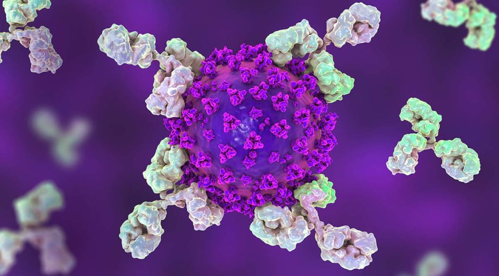
Loss of smell (olfaction) is a commonly reported symptom of COVID-19 infection. Recently, Bilinska, et al. set out to better understand the underlying mechanisms for loss of smell resulting from SARS-CoV-2 infection. In their research, they used in situ hybridization to investigate the expression of TMPRSS2, a SARS-CoV-2 viral entry protein in olfactory epithelium tissues of mice.

Why in situ hybridization?
In situ hybridization (ISH) is an important molecular method that is used for studying the function and regulation of genes. ISH uses hybridization probes to allow detection and localization of a specific nucleic acid sequence within a tissue sample or environmental sample. ISH can be used for population structure and morphology studies of microbes, and profiling gene expression in specific tissues.
There are several hybridization techniques currently used to perform ISH. In situ hybridization probes can be made from single- or double-stranded DNA, RNA, or even synthetic oligonucleotides. These hybridization probes are created by attaching a reporter molecule to a complementary strand of the target DNA or RNA. This process is called labeling.
When choosing a reporter molecule, it is important to decide which visualization method would be best for your research. When the hybridization probe is introduced to the sample, it binds to the nucleic acid of interest. Using the visualization method appropriate for the reporter molecule of choice, localization of the nucleic acid sequence of interest is possible.
DNA and RNA probes can be labeled with radioactive isotopes or non-radioactive molecules. Some examples of non-radioactive labels include biotin, digoxigenin, and fluorescent dye. Promega’s Riboprobe® Systems can be used for production of radio-labeled RNA, while RiboMAX™ Large Scale RNA Production Systems and RiboMAX™ Express products can be used for production of non-radiolabeled RNA. When fluorescence is used, the ISH technique is referred to as FISH. FISH is often used in studies where mapping chromosomes is important.
Using FISH to Study Expression of TMPRSS2 Protein
In viral research, in situ hybridization can be used to localize expression of a protein of interest in specific tissues. This technique was recently used by Bilinska, et al. in their search for expression of TMPRSS2, a SARS-CoV-2 viral entry protein, in olfactory epithelium tissues of mice (Figure 5 in the paper). A cDNA template containing sequence of the protein of interest was created. This cDNA was subcloned using the Promega pGEM-T Easy Vector Systems to make an easy to use template for in vitro transcription. RNA probes were then created using in vitro transcription and a digoxigenin-labeled nucleotide mix. Tissues were hybridized with the digoxigenin-labeled RNA probes and visualized with anti-digoxigenin antibodies and a chromogenic substrate. From this, the researchers were able to determine which cells in the olfactory epithelium may be targeted by TMPRSS2, as shown in Figure 1 of the paper. Bilinska, et al. conclude that the loss of sense of smell that has been reported in many people who test positive for COVID-19 may be caused by damage to sustentacular cells, which are responsible for supporting the sense of smell and metabolism of inhaled chemicals.
Learn more about our products to support Viral Research, Vaccine and Therapeutic Development.
Related Posts
Latest posts by Leah Cronan (see all)
- Mass Spec for Glycosylation Analysis of SARS-CoV-2 Proteins Implicated in Host-Cell Entry - November 10, 2020
- Proteomics from a Different Point of View: Introducing ProAlanase, the Newest Mass-Spec Grade Protease from Promega - August 7, 2020
- Go fISH! Using in situ Hybridization to Search for Expression of a SARS-CoV-2 Viral Entry Protein - July 10, 2020
