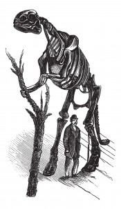
Brachylophosaurus was a mid-sized member of the hadrosaurid family of dinosaurs living about 78 million years ago, and is known from several skeletons and bonebed material from the Judith River Formation of Montana and the Oldman Formation of Alberta. Recent fossil evidence indicates structures similar to blood vessels in location and morphology, have been recovered after demineralization of multiple dinosaur cortical bone fragments from multiple specimens, some of which are as old as 80 Ma. These structures were hypothesized to be either endogenous to the bone (i.e., of vascular origin) or the result of biofilm colonizing the empty network after degradation of original organic components (i.e., bacterial, slime mold or fungal in origin). Cleland et al. (1) tested the hypothesis that these structures are endogenous and thus retain proteins in common with extant archosaur blood vessels that can be detected with high-resolution mass spectrometry and confirmed by immunofluorescence.
To optimize the recovery of endogenous proteins that are of lower abundance, but which may be more informative than collagen in living tissues, vessels were isolated by first demineralizing cortical bone from the tibia of a B. canadensis specimen for extraction rather than using whole bone extracts, which, in extant vertebrates, are dominated by collagen. Because birds are the closest living relatives of dinosaurs, blood vessels were also isolated from extant, demineralized ostrich and chicken bones, then the collagen I matrix was removed through specific collagenase digestion. Ostrich vessels were used as a positive control in immunohistochemical analyses, and ostrich and chicken vessels were used as positive controls for mass spec analysis.
Previous analyses of dinosaur peptides and soft tissues have been criticized as potentially arising from unknown contamination. Extensive measures were taken to minimize potential sources of contamination (e.g., using separate laboratories for preparation of extant and B. canadensis protein extracts and vessels; analyzing dinosaur peptides on instruments that had never been exposed to any avian proteins; employing extensive personal protective equipment to reduce human contamination that included nitrile gloves, lab coats, hair coverings, laminar flow hoods, and a dedicated “ancient” processing room).
Are you looking for proteases to use in your research?
Explore our portfolio of proteases today.
For mass spec analysis, protein pellets were resuspended in 50 mM ammonium bicarbonate, reduced for 1 h at 37 °C with 10 mM dithiothreitol and then alkylated for 1 h in the dark with 20 mM iodoacetamide. Proteins were digested with Promega modified trypsin overnight at 37 °C. After digestion, all samples were acidified with 1 μL of 100% formic acid. Peptides were concentrated by eluting from ZipTips with 50% acetonitrile 0.1% formic acid into 10 μL aliquots.
Using this process lead to the identification of cytoskeletal and nuclear peptide sequences from 10 different proteins. These proteins/peptides were not found in mass spectrometry analyses of the sediments extracted in tandem with the bone or in buffer controls. Nor were the peptides consistent with bacterial, slime mold, or fungal in origin. Furthermore, the proteins identified by mass spectrometry were validated by localized binding of antibodies raised against the same proteins.
1. Cleland, T. et.al (2015). Mass Spectrometry and Antibody-Based Characterization of Blood Vessels from Brachylophosaurus canadensis. J Proteome.Res. 14, 5252-62.

