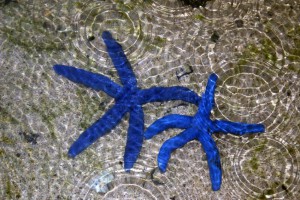
 A killer is lurking in the waters off the pacific coast. Silent and lethal, it leaves its decimated victims in tidal pools. They first began to appear in the early summer of 2013. Limp and curled, missing some or all of their limbs, the bodies were little more than globs of slimy tissue. They were hardly recognizable as what they once were—Sea Stars.
Typically found nestled in the cracks and crevices of tidal pools, sea stars (a.k.a., starfish) are the colorful iconic symbols of the beach. Starting in June of 2013 they began to die in alarming numbers up and down the pacific coast of North America, and not just in coastal waters, sea star populations in coastal aquariums also fell victim. The killer? A disease christened sea star wasting disease (SSWD).
Although sea star die offs attributed to SSWD have been reported before, the current outbreak differs not only in the breadth of its geographic range, but also by the unusually large number of species affected. These two things combine to make the current SSWD outbreak the largest known marine wildlife epizootic to date (1).
It was only after some, but not all, captive sea stars in coastal aquariums began to succumb to SSWD that scientists began to suspect that an infectious agent might be the cause. Sea star populations in coastal aquariums that used sand-filtered ocean water began dying, while sea star populations in aquariums that treated their ocean water with ultraviolet (UV) light remained healthy. It appeared that whatever caused SSWD was small enough to make it through sand filtration and was susceptible to UV treatment.
A team of scientists led by microbial oceanographer Ian Hewson sampled microorganisms from healthy and diseased individuals. When the microbial survey showed no major differences between the two populations, the scientists turned their suspicions to viruses. To test the idea that the infectious agent was a virus, they injected healthy sea stars with tissues from diseased individuals. Within three weeks of injection, the once health sea stars began to show symptoms of SSWD. However, if the tissue from infected sea star was heat treated to kill viruses and then injected, the recipient sea stars remained healthy. Scientist next isolated virus-sized particles from the symptomatic sea stars and injected this material into a new set of healthy sea stars. This second set of inoculated sea stars also showed signs of SSWD within three weeks, while those injected with a heat-treated sample of the isolated inoculate remained healthy (1).
Using metaviromic analysis, the team found a higher level of parvovirus-like sequences in samples from SSWD-affected sea stars than in those of health sea stars. Using all the viral metagenomes obtained, they were able to assemble an almost complete densovirus, which they named “sea star-associated densovirus” (SSaDV; 1).
As is true more often than not in science, the identification of SSaDV as a probable cause of the deadly disease afflicting the sea star populations of the Pacific coast is only the first piece of what is most likely a complicated puzzle. There are a number of questions as of yet unanswered. For example, Hewson’s team has found traces of densovirus in museum specimens that were collected over 72 years ago. This is not a new or exotic virus that has only recently appeared in these creatures, so why has the virus roared through the population with such a vengeance in the last year and a half? Hopefully as we learn more about SSWD scientist will eventually be able to predict, or perhaps even stop, an outbreak.
There is some good news coming out of recent tidal pool surveys. In November, researchers at Point Conception in Santa Cruz, CA found hundreds of thumbnail-sized sea star babies. In other areas where populations were all but whipped out, large numbers of juvenile sea stars have been spotted. For now, it seems, the killer is retreating.
Reference
- Hewson, I. et al. (2014) Densovirus associated with sea-star wasting disease and mass mortality. Proc. Natl. Acad. Sci. 48, 17278–83.
