For those of us entering the world of cell-based assays from a classical or molecular genetics background, the world of cell culture can be daunting. Yet to truly understand how the genetic mutation behind a particular phenotype works, we need to look at the biochemistry and cell biology where it all occurs: the cell.
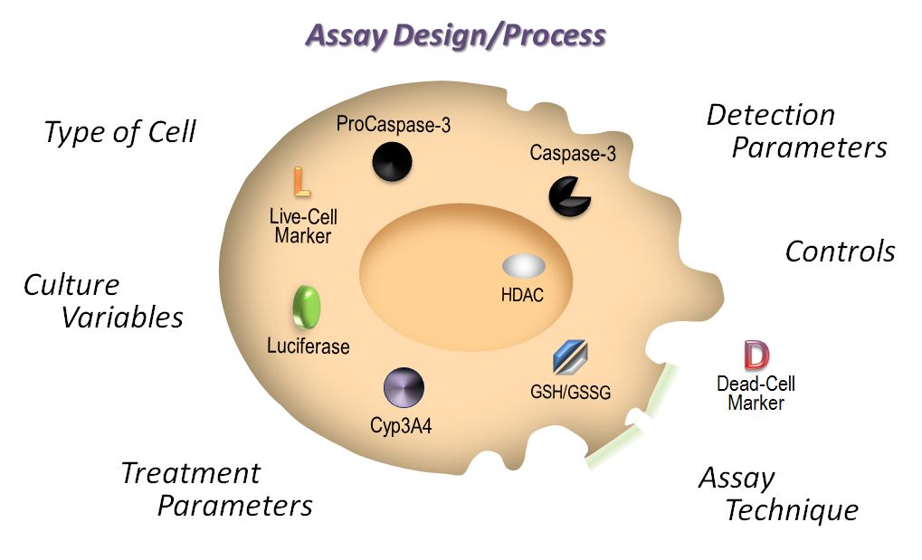
This series of blogs will cover several topics to consider when designing your cell-based assays. In this first installment, we discuss the basics of choosing the cell type for your assay.
When deciding what cells to use for your assay three basic things are essential. The cells must:
- be amenable to the assay,
- faithfully represent the system, and
- express the necessary factors and signaling intermediates
Among commonly used cells, fibroblasts (HEK293, Cos cells), are an easy, amenable experimental system; however, they may not be the most relevant to the question you are asking. Cancer lines (HepG2 and PC-3 cells) maybe more relevant and are generally easy to use, but remember that a cancer line is just that, a cancer line. It can contain mutations that might affect your experimental outcome. For instance, the cancer cell line MCF-7 lacks a functional caspase-3 gene product, so if you are studying apoptosis using a DEVD substrate, you may underestimate it. Primary cells (HUVECs and hepatocytes) may afford the most accurate picture of the typical in vivo situation, but they can be difficult to grow and transfect.
The cell type that you choose will affect how you design your assay. For example, if you are performing a cell viability assay that uses a metabolite (e.g., ATP), you will need to remember that cell lines generally have a higher metabolic rate than primary cells. Additionally, primary cells in culture have a higher spontaneous rate of death, so if you are performing a cytotoxicity assay (e.g., LDH release), you can expect a higher background from an assay performed using primary cells.
Other factors that can influence your assay results include:
- treatment choice, duration and concentration,
- assay incubation time, and
- seeding density
Different cell types have different susceptibility to injury or test compounds that are cytotoxic or cause apoptosis. The same agent that induces apoptosis in one cell line may have no effect on a different cell line, or an agent that does induce apoptosis in two different cell lines may do so with a radically different time courses. So, consulting the literature for recommended assay protocols or conducting preliminary experiments to determine optimal conditions for your system is recommended. Perform an initial dose response curve to identify the appropriate drug concentration or stimulus intensity to induce apoptosis/cytotoxicity in the cell or tissue of interest, and examine the biochemical event over a sufficiently broad time period with appropriate time increments to capture peak activity and minimize erroneous conclusions based on ill-timed assay points.
Factors such as cell density, passage number and the density to which the parental stock culture was grown can affect cell physiology, and subsequently, may affect the ability to respond to a stimulus. To control for these kinds of effects, be sure to use well established culture procedures and consistent cell numbers to minimize these variables.
These are a few of the basic things to consider when choosing your cell type for a cell-based assay. Below are some resources to assist you with your assay design and cell choice.
Next installment: More about Basic Cell Culture Conditions. Stay tuned!
Multimedia:
Culture Preparation and Plating
Articles:
Timing Your Apoptosis Assays
Multiplexing Cell-Based Assays: Get More Biologically Relevant Data
Choosing the Right Cell-Based Assay for Your Research
Michele Arduengo
Latest posts by Michele Arduengo (see all)
- An Unexpected Role for RNA Methylation in Mitosis Leads to New Understanding of Neurodevelopmental Disorders - March 27, 2025
- Unlocking the Secrets of ADP-Ribosylation with Arg-C Ultra Protease, a Key Enzyme for Studying Ester-Linked Protein Modifications - November 13, 2024
- Exploring the Respiratory Virus Landscape: Pre-Pandemic Data and Pandemic Preparedness - October 29, 2024


Hi Michelle,
Very nice post. Makes me miss my cell culture facility. (sniff, sniff)
I am paying “blog calls” to each @scio12 attendee to say “Hi” and give your blog a shoutout on twitter (I’m @sciencegoddess). I look forward to meeting you in January!
Thanks for visiting. I love the idea of visiting the blogs of attendees at scio12. I may steal your idea. Glad you like the blog. We try to be helpful around here.