Understanding and Combating Legionella in Water Systems with Viability PCR

Water plays a vital role in countless aspects of daily life—drinking, cooling, recreation and more. However, the same systems that deliver these benefits can also harbor Legionella, a waterborne bacteria responsible for Legionnaires’ disease, a severe form of pneumonia1. Legionella thrives in stagnant aquatic environments, many of which are human-made and common in modern infrastructure, like in cooling towers, hot tubs and complex building water systems. In this blog, we explore the risks posed by Legionella, the limitations of traditional detection methods and how advanced tools at Promega are transforming water safety monitoring. 

Legionella Detection: Challenges and Emerging Solutions 

In 1976, a pneumonia outbreak at a Philadelphia convention led to the discovery of Legionella pneumophila, the bacterium behind Legionnaires’ disease. The pathogen was traced to aerosolized water from the air conditioning system, infecting 182 people and causing 29 deaths2. This incident highlighted the far-reaching consequences of contaminated water systems, which threaten public health and impose significant financial and operational burdens on businesses and institutions. Cooling towers, decorative water features, and even hospital water systems can become sources of infection if proper precautions aren’t taken. For vulnerable populations—including older adults and those with weakened immune systems—exposure to Legionella can lead to life-threatening illness. 

In addition to being time-intensive, culture-based detection can be unreliable in mixed microbial populations. Faster-growing, non-Legionella bacteria can overgrow culture plates, obscuring the presence of Legionella and leading to underreporting of contamination3. Culture methods are also unable to detect viable-but-non-culturable (VBNC) cells—bacteria that remain infectious but cannot grow under standard laboratory conditions. These hidden risks are a critical gap in traditional culture methods and highlight the need for more comprehensive detection approaches. 

To address the limitations of culture-based methods, many facilities have turned to polymerase chain reaction (PCR) for faster and more sensitive results. PCR can detect Legionella DNA in as little as a few hours, offering same-day conformation of water quality. However, traditional PCR methods don’t differentiate between live, infectious bacteria and dead cells, which can lead to false positives and complicate risk assessments. This limitation underscores the importance of innovations like a viability PCR, which offers enhanced accuracy by focusing on live-cell detection. 

Advanced Detection with Viability PCR  

To overcome the challenges of traditional detection methods, Promega has developed a novel viability PCR workflow that sets a new standard for Legionella testing. At the heart of this solution is a dye that selectively inactivates DNA from dead cells, allowing for the detection of live versus dead Legionella cells. This innovation eliminates false positives caused by residual DNA from non-viable bacteria, providing highly accurate and informed results. 

Workflow for Legionella testing with PCR Viability kits at Promega. Top section includes each step in the workflow: gathering the sample, sample neutralization, DNA purification, and real-time PCR. Below the workflow image is time for each step and a description of each step.
Viability PCR Workflow.

The Viability PCR workflow integrates seamlessly into water testing protocols, offering flexibility for both manual and automated DNA extraction methods. The Wizard® PureWater Kit provides an efficient, hands-on solution for labs processing smaller sample volumes, while the Maxwell® RSC PureWater System delivers automated, high-throughput DNA purification for larger-scale operations. These options allow water quality professionals, public health laboratories, and environmental monitoring teams to customize workflows based on the unique needs of their facility. 

By combining selective viability detection with multiplex qPCR technology, the Viability PCR solution enables the simultaneous identification of multiple Legionella species, including Legionella pneumophila and its most pathogenic serogroup, SG1, which are linked to outbreaks of Legionnaire’s disease. This comprehensive approach ensures that water safety monitoring is not only faster but also more precise, providing institutions with the tools they need to protect public health. 


To learn more about Legionella detection solutions at Promega, check out this page.


References

  1. “Legionella (Legionnaires’ Disease and Pontiac Fever): About.” Centers for Disease Control and Prevention, www.cdc.gov/legionella/about/index.html. Accessed 16 Jan. 2025. ↩︎
  2. “Legionella Pneumophila.” National Center for Biotechnology Information, U.S. National Library of Medicine, 23 July 2018, www.ncbi.nlm.nih.gov/books/NBK430807/. Accessed 16 Jan. 2025. ↩︎
  3. Miskowski, Diane. “An Overview of Legionella.” EMSL Analytical, Inc., www.legionellatesting.com/legionella-article/?utm_source=chatgpt.com. Accessed 16 Jan. 2025. ↩︎

Our Maxwell® Travels from Spain to Antarctica to Help Stop the Avian Flu Virus

In January 2024, Antonio Alcamí and Ángela Vázquez, virologists from the Severo Ochoa Centre for Molecular Biology, landed in Antarctica to study the avian flu virus. They embarked on a journey to monitor 17,000 penguins as part of their efforts to study the virus and prevent its spread. Our Maxwell® RSC 48 was delivered to extract nucleic acids from the samples, which are set to be analyzed using qPCR.

Continue reading “Our Maxwell® Travels from Spain to Antarctica to Help Stop the Avian Flu Virus”

An Introduction to Lyophilization: Process, Benefits & Possibilities

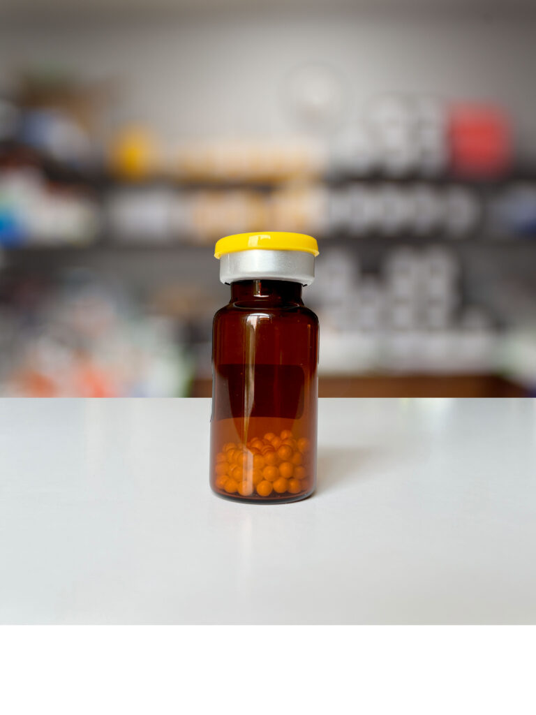
Amber glass bottle filled with lyophilized beads sitting on a lab bench.

Lyophilization is a process designed to remove water from a sample or product through a controlled freezing and vacuum application. The method leverages the triple point of water, where solid, liquid, and gas phases coexist under specific temperature and pressure conditions. The result is a room temperature stable product that is much lighter than the original sample or product.

Continue reading “An Introduction to Lyophilization: Process, Benefits & Possibilities”

Promega qPCR Grant Series #3: Immunotherapy Researcher, Dr. Sabrina Alves dos Reis 

Professional headshot image of Dr. Sabrina Alves dos Reis, subject of the blog post
Sabrina Alves dos Reis

In our third and final installment of the Promega qPCR Grant Recipient blog series, we highlight Dr. Sabrina Alves dos Reis, a trained immunotherapy researcher. Her work has focused on developing tools for more accessible cancer therapies using CAR-T cells. Here, we explore Dr. Alves dos Reis’ academic and scientific journeys, highlight influential mentorship and foreshadow her plans for the Promega qPCR grant funds. 

Dr. Alves dos Reis’ career began with a strong affinity for biology. As an undergraduate student, she pursued a degree in biological science, where she developed a foundational understanding for designing and developing research projects. As her passion for science heightened, she decided to continue her journey in science, culminating in a PhD at the Fundação Oswaldo Cruz Institute in Rio de Janeiro, Brazil. Her research projects focused on the unexplored territory of adipose tissue as a site for Mycobacterium leprae—or leprosy bacillus—infection. She mentioned that this work piqued her curiosity for improving immunotherapies and laid the foundation for her future in cancer research.  

Continue reading “Promega qPCR Grant Series #3: Immunotherapy Researcher, Dr. Sabrina Alves dos Reis “

Promega qPCR Grant Series #2: Molecular Biologist, Laura Leighton

Our second installment of the Promega qPCR Grant Recipient blog series highlights Dr. Laura Leighton, a trained molecular biologist and postdoctoral researcher at the Australian Institute for Bioengineering and Nanotechnology. Leighton’s scientific journey features a passion for molecular biology and problem-solving. Her path has been illuminated by mentorship, relationships with fellow scientists and a commitment to creativity in overcoming challenges. Here, we explore her scientific journey, reflect on research lessons and foreshadow her plans for the Promega qPCR grant funds.

Dr. Laura Leighton grew up in a rural area in Far North Queensland, Australia, where she spent her early life exploring critters on the family farm. Her upbringing was infused with a deep connection to the environment, from raising tadpoles in wading pools to observing wildlife and witnessing food grow firsthand. Observing the biology around her ultimately piqued her interest in science from a young age. She then began her academic journey in 2011 at the University of Queensland, Australia. She studied biology while participating in a program for future researchers, which led her to undergraduate research work in several research labs.  She dabbled in many research avenues in order to narrow in on her scientific interests all while adding different research tools to her repertoire.

After serving as a research assistant in Dr. Timothy Bredy’s lab, she decided to continue work in this lab and pursue a PhD in molecular biology. During her PhD, Leighton worked on several projects from cephalopod mRNA interference to neurological wiring in mice. The common thread in these projects is Leighton’s passion for the puzzles of molecular biology:

“I also love molecular engineering and the modularity of molecular parts. There’s something really special about stringing together sequence in a DNA editor, then seeing it come to life in a cell,” she says.

Continue reading “Promega qPCR Grant Series #2: Molecular Biologist, Laura Leighton”

Promega qPCR Grant Series #1: Marine Plant Ecologist, Dr. Agustín Moreira-Saporiti

Dr. Agustín Moreira-Saporiti is a postdoctoral researcher at the Marine Biological Laboratory and is studying flowering processes in marine seagrass

Marine seagrasses are submerged flowering plants that form essential underwater meadows, fostering diverse ecosystems and providing a habitat for marine life. Our first Promega qPCR Grant winner and marine ecologist, Dr. Agustín Moreira-Saporiti, plans to continue adding to a fascinating body of work aimed at understanding flowering in marine seagrasses.

Dr. Moreira-Saporiti began his journey into marine plant ecology at the University of Vigo, Spain, where he earned a bachelor’s degree in marine sciences. He then went on to complete a master’s degree at the University of Bremen (Germany) where his thesis focused the ecology of seagrasses in Zanzibar, Tanzania. His passion for marine botany led him down a deeper exploration of marine plants, unraveling the intricate web of ecosystem processes within seagrasses.

Continue reading “Promega qPCR Grant Series #1: Marine Plant Ecologist, Dr. Agustín Moreira-Saporiti”

No Horsin’ around with Halal Meat Authentication


Today’s blog is written by guest blogger, Sameer Moorji, Director, Applied Markets.  

People’s diets are frequently influenced by a wide range of variables; with environment, socioeconomic status, religion, and culture being a few of the key influencers. The Muslim community serves as one illustration of how culture and religion can hold influence over people’s eating habits.

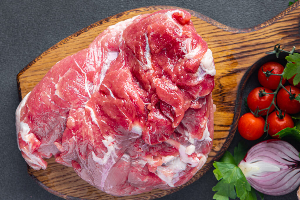
Halal meat on cutting board

Muslims, who adhere to Islamic teachings derived from the Qur’an, frequently base dietary choices on a food’s halal status, whether it is permissible to consume, or haram status, forbidden to consume. With the population of Muslims expected to expand from 1.6 billion in 2010 to 2.2 billion by 2030, the demand for halal products is anticipated to surge (2).

By 2030, the global halal meat market is projected to reach over $300 billion dollars, with Asia-Pacific and the Middle East regions being the largest consumers and producers of halal meat products (3). Furthermore, increasing awareness and popularity of halal meat among non-Muslim consumers, as well as strengthening preference for ethical and high-quality meat, are all contributing to demand.  

Continue reading “No Horsin’ around with Halal Meat Authentication”

Have No Fear, qPCR Is Here: How qPCR can help identify food contamination

Foodborne disease affects almost 1 in 10 people around the world annually, and continuously presents a serious public health issue (9).

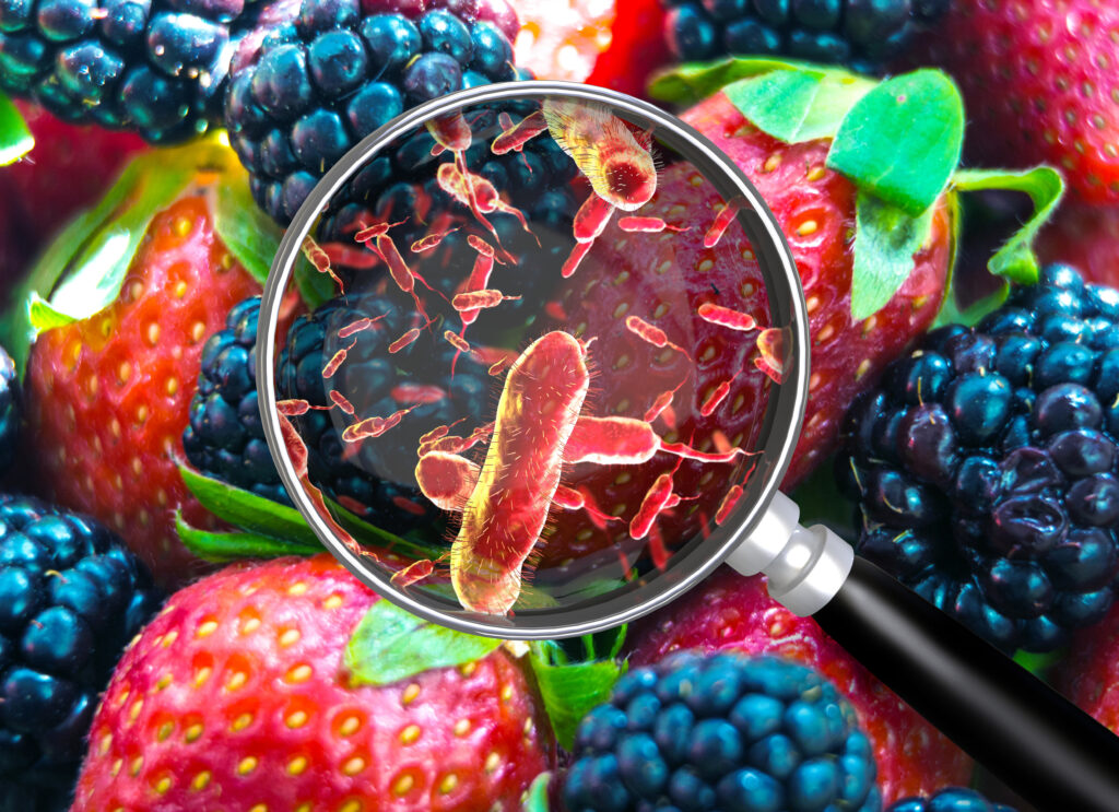
Food Contamination-Strawberries-Blueberries-Magnifying glass
Food Contamination is common and can be seen in a variety of forms and food products.

More than 200 diseases have evolved from consuming food contaminated by bacteria, viruses, parasites, and chemical substances, resulting in extensive increases in global disease and mortality rates (9). With this, foodborne pathogens cause a major strain on health-care systems; as these diseases induce a variety of different illnesses characterized by a multitude of symptoms including gastrointestinal, neurological, gynecological, and immunological (9,2).

But why is food contamination increasing?

New challenges, in addition to established food contamination hazards, only serve to compound and increase food contamination risks. Food is vulnerable to contamination at any point between farm and table—during production, processing, delivery, or preparation. Here are a few possible causes of contamination at each point in the chain (2):

  • Production: Infected animal biproducts, acquired toxins from predation and consumption of other sick animals, or pollutants of water, soil, and/or air.
  • Processing: Contaminated water for cleaning or ice. Germs on animals or on the production line.
  • Delivery: Bacterial growth due to uncontrolled temperatures or unclean mode of transport.
  • Preparation: Raw food contamination, cross-contamination, unclean work environments, or sick people near food.

Further emerging challenges include, more complex food movement, a consequence of changes in production and supply of imported food and international trade. This generates more contamination opportunities and transports infected products to other countries and consumers. Conjointly, changes in consumer preferences, and emerging bacteria, toxins, and antimicrobial resistance evolve, and are constantly changing the game for food contamination (1,9).

Hence, versatile tests that can identify foodborne illnesses in a rapid, versatile, and reliable way, are top priority.

Continue reading “Have No Fear, qPCR Is Here: How qPCR can help identify food contamination”

qPCR: The Very Basics

Real-Time (or quantitative, qPCR) monitors PCR amplification as it happens and allows you to measure starting material in your reaction.
qPCR monitors amplification in real and allows you to measure starting material.

For those of us well versed in traditional, end-point PCR, wrapping our minds and methods around real-time or quantitative (qPCR) can be challenging. Here at Promega Connections, we are beginning a series of blogs designed to explain how qPCR works, things to consider when setting up and performing qPCR experiments, and what to look for in your results.

First, to get our bearings, let’s contrast traditional end-point PCR with qPCR.

End-Point PCRqPCR
Visualizes by agarose gel the amplified product AFTER it is produced (the end-point)Visualizes amplification as it happens (in real time) via a detection instrument
Does not precisely measure the starting DNA or RNAMeasures how many copies of DNA or RNA you started with (quantitative = qPCR)
Less expensive; no special instruments requiredMore expensive; requires special instrumentation
Basic molecular biology techniqueRequires slightly more technical prowess

Quantitative PCR (qPCR) can be used to answer the same experimental questions as traditional end-point PCR: Detecting polymorphisms in DNA, amplifying low-abundance sequences for cloning or analysis, pathogen detection and others. However, the ability to observe amplification in real-time and detect the number of copies in the starting material can quantitate gene expression, measure DNA damage, and quantitate viral load in a sample and other applications.

Anytime that you are performing a reaction where something is copied over and over in an exponential fashion, contaminants are just as likely to be copied as the desired input. Quantitative PCR is subject to the same contamination concerns as end-point PCR, but those concerns are magnified because the technique is so sensitive. Avoiding contamination is paramount for generating qPCR results that you can trust.

  1. Use aerosol-resistant pipette tips, and have designated pipettors and tips for pre- and post-amplification steps.
  2. Wear gloves and change them frequently.
  3. Have designated areas for pre- and post-amplification work.
  4. Use reaction “master mixes” to minimize variability. A master mix is a ready-to-use mixture of your reaction components (excluding primers and sample) that you create for multiple reactions. Because you are pipetting larger volumes to make the reaction master mix, and all of your reactions are getting their components from the same master mix, you are reducing variability from reaction to reaction.
  5. Dispense your primers into aliquots to minimize freeze-thaw cycles and the opportunity to introduce contaminants into a primer stock.

These are very basic tips that are common to both end-point and qPCR, but if you get these right, you are off to a good start no matter what your experimental goals are.

If you are looking for more information regarding qPCR, watch this supplementary video below.


We’re committed to supporting scientists who are using molecular biology to make a difference. Learn more about our qPCR Grant program.  


Are you looking for more in-depth information about qPCR? Check out our qPCR and RT-qPCR Guide!


Related Posts

Optimizing PCR: One Scientist’s Not So Fond Memories

primer_tubesThe first time I performed PCR was in 1992. I was finishing my Bachelors in Genetics and had an independent study project in a population genetics laboratory. My task was to try using a new technique, RAPD PCR, to distinguish clonal populations of the sea anemone, Metridium senile. These creatures can reproduce both sexually and asexually, which can make population genetics studies challenging. My professor was looking for a relatively simple method to identify individuals who were genetically identical (i.e., potential clones).

PCR was still in its infancy. No one in my lab had ever tried it before, and the department had one thermal cycler, which was located in a building across the street. We had a paper describing RAPD PCR for population work, so we ordered primers and Taq DNA polymerase and set about grinding up bits of frozen sea anemone to isolate the DNA. [The grinding process had to be done using a mortar and pestle seated in a bath of liquid nitrogen because the tissue had to remain frozen. If it thawed it became a disgusting mass of goo that was useless—but that is a topic for a different blog.] Since I had never done any of the procedures before, my professor and I assembled the first set of reactions together. When we ran our results on a gel, we had all sorts of bands—just what he was hoping to see. Unfortunately, we realized that we had added 10X more Taq DNA polymerase than we should have used. I repeated the amplification with the correct amount of Taq polymerase, and I saw nothing. Continue reading “Optimizing PCR: One Scientist’s Not So Fond Memories”