Weird samples? Contact Tech Serv to find the right DNA purification kit for you.

“Dear Tech Serv,
We would like to detect DNA collected from swabs rubbed on the inside thighs of frogs. What would be the best DNA extraction kit to use for this?”

“Hi Tech Serv,
I need to find out a suitable kit for extracting DNA from bird fecal samples. Can I use ReliaPrep™ gDNA Tissue Miniprep System for that?”

These are just some examples of unconventional sample type inquiries that the Promega Technical Services Team receives regularly from scientists around the world. Many of these inquiries land in the hands of Technical Services Scientist, Paraj Mandrekar (a.k.a. “sample type guru”).

Continue reading “Weird samples? Contact Tech Serv to find the right DNA purification kit for you.”

Better NGS Size Selection

One of the most critical parts of a Next Generation Sequencing (NGS) workflow is library preparation and nearly all NGS library preparation methods use some type of size-selective purification. This process involves removing unwanted fragment sizes that will interfere with downstream library preparation steps, sequencing or analysis.

Different applications may involve removing undesired enzymes and buffers or removal of nucleotides, primers and adapters for NGS library or PCR sample cleanup. In dual size selection methods, large and small DNA fragments are removed to ensure optimal library sizing prior to final sequencing. In all cases, accurate size selection is key to obtaining optimal downstream performance and NGS sequencing results.

Current methods and chemistries for the purposes listed above have been in use for several years; however, they are utilized at the cost of performance and ease-of-use. Many library preparation methods involve serial purifications which can result in a loss of DNA. Current methods can result in as much as 20-30% loss with each purification step. Ultimately this may necessitate greater starting material, which may not be possible with limited, precious samples, or the incorporation of more PCR cycles which can result in sequencing bias. Sample-to-sample reproducibility is a daily challenge that is also regularly cited as an area for improvement in size-selection.

Continue reading “Better NGS Size Selection”

How Do I Choose the Right GoTaq® Product to Suit My Needs for EndPoint PCR?

We offer a wide array of GoTaq® DNA Polymerases, Buffers and Master Mixes, so we frequently answer questions about which product would best suit a researcher’s needs. On the Taq Polymerase Page, you can filter the products by clicking the categories on the left hand side of the page to narrow down your search. Here are some guidelines to help you select the match that will best suit your PCR application. Continue reading “How Do I Choose the Right GoTaq® Product to Suit My Needs for EndPoint PCR?”

Six (and a Half) Reasons to Quantitate Your DNA

Knowing how much DNA you have is fundamental to successful experiments. Without a firm number in which you are confident, the DNA input for subsequent experiments can lead you astray. Below are six reasons why you should quantitate your DNA.

6. Saving time by knowing what you have rather than repeating experiments. If you don’t quantitate your DNA, how certain can you be that the same amount of DNA is consistently added? Always using the same volume for every experiment does not guarantee the same DNA amount goes into the assay. Each time there is a new purified DNA sample, the chances that you have the same quantity as before are lessened. Consequently, without knowing the DNA concentration of the sample you are using, the amount of input DNA cannot be guaranteed and experiments may have to be repeated.

Continue reading “Six (and a Half) Reasons to Quantitate Your DNA”

Three Factors That Can Hurt Your Assay Results

4621CA

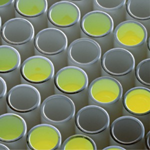
Each luminescent assay plate represents precious time, effort and resources. Did you know that there are three things about your detection instrument that can impact how much useful information you get from each plate?  Instruments with poor sensitivity may cause you to miss low-level samples that could be the “hit” you are looking for.  Instruments with a narrow detection range limit the accuracy or reproducibility you needed to repeat your work.  Finally, instruments that let the signal from bright wells spill into adjacent wells allow crosstalk to occur and skew experimental results, costing you time and leading to failed or repeated experiments.

Continue reading “Three Factors That Can Hurt Your Assay Results”

The Cell Line Identity Crisis

29981164-Whistle

If you work with cell lines you may have paid attention to the dramatic headline published last month in the online journal STAT, Thousands of studies used the wrong cells, and journals are doing nothing.” In their column The Watchdogs (“Keeping an eye on misconduct, fraud, and scientific integrity”), Ivan Oransky and Adam Marcus call out the fact that scientists continue to publish research using cell lines that are contaminated or misidentified. Recent estimates have found that the percentage of misidentified cell lines used by scientists is as high as 20 to 36. The blame here is being placed on the peer reviewed journals for not blowing the whistle. The authors call for journals to put some “kind of disclaimer on the thousands of studies affected.”

This is not a new claim. The continuing problem of cell line misidentification, of lack of authentication, has been covered before in various channels. It’s easy to find news publicizing yet another retracted publication. In May 2015 the journal Nature required authors of all submitted manuscripts to confirm the identity of cell lines used in their studies and provide details about the source and testing of their cell lines.

Continue reading “The Cell Line Identity Crisis”

Improving the Success of Your Transfection

12150558-plasmid_with_cell_membrane3

Not every lab has a tried and true transfection protocol that can be used by all lab members. Few researchers will use the same cell type and same construct to generate data. Many times, a scientist may need to transfect different constructs or even different molecules (e.g., short-interfering RNA [siRNA]) into the same cell line, or test a single construct in different cultured cell lines. One construct could be easily transfected into several different cell lines or a transfection protocol may work for several different constructs. However, some cells like primary cells can be difficult to transfect and some nucleic acids will need to be optimized for successful transfection. Here are some tips that may help you improve your transfection success.

Transfect healthy, actively dividing cells at a consistent cell density. Cells should be at a low passage number and 50–80% confluent when transfected. Using the same cell density reduces variability for replicates. Keep cells Mycoplasma-free to ensure optimal growth.

Transfect using high-quality DNA. Transfection-quality DNA is free from protein, RNA and chemical contamination with an A260/A280 ratio of 1.7–1.9. Prepare purified DNA in sterile water or TE buffer at a final concentration of 0.2–1mg/ml.

Continue reading “Improving the Success of Your Transfection”

Ten Validation Tips You Need to Know

Forensic lab validations can be intimidating, so Promega Technical Services Support and Validation teams shared these tips for making the process go more smoothly.

  1.  Prepare Your Lab. Make sure all of your all of your instrumentation (CEs, thermal cyclers, 7500s, centrifuges) and tools (pipettes, heat blocks) requiring calibration or maintenance are up to date.
  2.  Start with Fresh Reagents. Ensure you have all required reagents and that they are fresh before beginning your validation. This not only includes the chemistry being validated, but any preprocessing reagents or secondary reagents like, polymer, buffers, TE-4 or H2O.
  3. Develop a Plan. Before beginning a validation, take the time to create plate maps, calculate required reagent volumes, etc. This up-front planning may take some time initially, but will greatly improve your efficiency during testing.
  4. Create an Agenda. After a plan is developed, work through that plan and determine how and when samples will be created and run. Creating an agenda will hold you to a schedule for getting the testing done.
  5. Determine the Number of Samples Needed to Complete Your Validation. Look at your plan and see where samples can be used more than once.  The more a sample can be used, the less manipulation done to the sample and the more efficient you become.
  6. Select the Proper Samples for Your Validation. Samples should include those you know you’ll obtain results with be similar to the ones you’ll most likely be using, and your test samples should contain plenty of heterozygotes. When you are establishing important analysis parameters, like thresholds, poor sample choice may cause more problems and require troubleshooting after the chemistry is brought on-line.
  7. Perform a Fresh Quantitation of Your Samples. This will ensure the correct dilutions are prepared. Extracts that have been sitting for a long time may have evaporated or contain condensation, resulting in a different concentration than when first quantitated.
  8. Stay Organized. Keep the data generated in well-organized folders. Validations can contain a lot of samples, and keeping those data organized will help during the interpretation and report writing phase.
  9. Determine the Questions to Be Answered. While writing the report, determine the questions each study requires to be answered. Determining what specifically is required for each study will prevent you from calculating unnecessary data.  Do you need to calculate allele sizes of your reproducibility study samples when you showed precision with your ladder samples?
  10. Have fun! Remember, validations are not scary when approached in a methodical and logical fashion. You have been chosen to thoroughly test something that everyone in your laboratory will soon be using. Take pride in that responsibility and enjoy it.

Need more information about validation of DNA-typing products in the forensic laboratory? Check out the validation resources on the Promega web site for more information for the steps required to adopt a new product in your laboratory and the recommended steps that can help make your validation efforts less burdensome.

For Protein Complementation Assays, Design is Everything

Most, if not all, processes within a cell involve protein-protein interactions, and researchers are always looking for better tools to investigate and monitor these interactions. One such tool is the protein complementation assay (PCA). PCAs use  a reporter, like a luciferase or fluorescent protein, separated into two parts (A and B) that form an active reporter (AB) when brought together. Each part of the split reporter is attached to one of a pair of proteins (X and Y) forming X-A and Y-B. If X and Y interact, A and B are brought together to form the active enzyme (AB), creating a luminescent or fluorescent signal that can be measured. The readout from the PCA assay can help identify conditions or factors that drive the interaction together or apart.

A key consideration when splitting a reporter is to find a site that will allow the two parts to reform into an active enzyme, but not be so strongly attracted to each other that they self-associate and cause a signal, even in the absence of interaction between the primary proteins X and Y. This blog will briefly describe how NanoLuc® Luciferase was separated into large and small fragments (LgBiT and SmBiT) that were individually optimized to create the NanoBiT® Assay and show how the design assists in monitoring protein-protein interactions.

Continue reading “For Protein Complementation Assays, Design is Everything”

T-Vector Cloning: Answers to Frequently Asked Questions

Blue/White colony screening helps you pick only the colonies that have your insert.

Q: Can PCR products generated with GoTaq® DNA Polymerase be used to for T- vector cloning?

A: Yes. GoTaq® DNA Polymerase is a robust formulation of unmodified Taq Polymerase. GoTaq®DNA Polymerase lacks 3’ →5’ exonuclease activity (proof reading) and also displays non-template–dependent terminal transferase activity that adds a 3′ deoxyadenosine (dA) to product ends. As a result, PCR products amplified using GoTaq® DNA Polymerase will contain A-overhangs which makes it suitable for T-vector cloning.

We have successfully cloned PCR products generated using GoTaq® and GoTaq® Flexi DNA Polymerases into the pGEM®-T (Cat.# A3600), pGEM®-T Easy (Cat.# A1360) and pTARGET™ (Cat.# A1410) Vectors.

Q: Can GoTaq® Long PCR Master Mix be used for T-Vector Cloning?

A: Yes it can. GoTaq® Long PCR Master Mix utilizes recombinant Taq DNA polymerase as well as a small amount of a recombinant proofreading DNA polymerase. This 3´→5´ exonuclease activity (proof reading) enables amplification of long targets. Despite the presence of a small amount of 3´→5´ exonuclease activity, the GoTaq® Long PCR Master Mix generates PCR products that can be successfully ligated into the pGEM®-T Easy Vector System.

We have demonstrated that GoTaq® Long PCR Master Mix successfully generated DNA fragments that could be ligated into pGEM®-T Easy Vector System without an A-tailing procedure, and with ligation efficiencies similar to those observed with the GoTaq® Green Master Mix.

For details refer to Truman, A., Hook, B. and Wieczorek, D. Using GoTaq® Long PCR Master Mix for T-Vector Cloning.

Tip: For cloning blunt-ended PCR fragments into T-vectors, use the A-tailing protocol discussed in the pGEM®-T and pGEM®-T Easy Technical Manual #TM042.

Q: How do I prepare PCR products for ligation? What products can be used to purify the DNA?

Continue reading “T-Vector Cloning: Answers to Frequently Asked Questions”