A New Edge in Bisulfite Conversion

methyledge_featureproduct_280x140

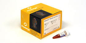
Aberrant methylation events have significant impacts in terms of incidence of cancer and development disregulation. Researchers studying DNA methylation are often working with DNA from “difficult” tissues such as formalin-fixed, paraffin embedded tissues, which characteristically yield DNA that is more fragmented than that purified from fresh tissue. Traditional methods for bisulfite conversion involve a long protocol, harsh chemicals, and generally yield highly fragmented DNA. The DNA fragmentation may significantly impact the utility of the converted DNA in downstream applications such as bisulfite-specific PCR or bisulfite sequencing.

An ideal bisulfite conversion system enables complete conversion of a DNA sample in a short period of time, provides high yield of DNA, minimally fragments the DNA, works on a wide range of input DNA amounts (from a wide variety of sample types), and, while we’re at it, is easy to use and to store. Whew! That’s quite the list.

Continue reading “A New Edge in Bisulfite Conversion”

“Fingerprinting” Your Cell Lines

Working in the laboratoryResearchers working with immortalized cell lines would readily agree when I state that it is almost impossible to look at cells under the microscope and identify them by name. There are phenotypic traits, however they do change with change in media composition, passage number and in response to growth factors. I remember the pretty arborizations my neuroblastoma cell line SH-SY5Y exhibited in response to nerve growth factor treatment. Thus physical appearance is not a distinguishing feature. Currently, in many labs, researchers typically use more than one cell line, and more than likely, share the same lab space to passage cells and the same incubator to grow the cells. In such scenarios, it is not difficult to imagine that cell lines might get mislabeled or cross-contaminated. For example HeLa cells, one of the fastest growing cell lines have been shown to invade and overtake other cell lines.

Misidentification of cell lines has deep and severe implications. A review of cell lines used to study esophageal adenocarcinoma found that a large number of the cell lines were actually derived from lung or gastric cancers. Unfortunately, by the time this error was discovered, data from these cell line studies were already being used for clinical trials and other advanced studies and publications. Moreover, the cell lines were being to screen and design and test specific cancer drugs which ended up in flawed clinical trials. Continue reading ““Fingerprinting” Your Cell Lines”

DNA Purification, Quantitation and Analysis Explained

WebinarsYesterday I listened in on the Webinar “Getting the Most Out of Your DNA Analysis from Purification to Downstream Assays”, presented by Eric Vincent–a Product Manager in the Promega Genomics group.

This is the webinar for you if you have ever wondered about the relative advantages and disadvantages of the many methods available for DNA purification, quantitation and analysis, or if you are comparing options for low- to high-throughput DNA purification. Eric presents a clear analyses of each of the steps in a basic DNA workflow: Purification, Quantitation, Quality Determination, and Downstream Analysis, providing key considerations and detailing the potential limitations of the methods commonly used at each step.

The DNA purification method chosen has an affect on the quality and integrity of the DNA isolated, and can therefore affect performance in downstream assays. Accuracy of quantitation also affects success, and the various downstream assays themselves (such as end-point PCR, qPCR, and sequencing) each have different sensitivities to factors such as DNA yield, quality, and integrity, and the presence of inhibitors. Continue reading “DNA Purification, Quantitation and Analysis Explained”

Choosing the Right Reverse Transcriptase for Your Project

There are a lot of choices when it comes to reverse transcriptases.  Choosing the correct one for your cDNA synthesis and RT-PCR project is important.    Here are a few questions that will lead you to right RT for your application: Continue reading “Choosing the Right Reverse Transcriptase for Your Project”

Methods for Determining DNA Yield and Concentration

Determining DNA Yield and Purity

This post is provided as a general introduction to common laboratory methods for determining the yield and purity of purified DNA samples. DNA yield can be assessed using various methods including absorbance (optical density), agarose gel electrophoresis, or use of fluorescent DNA-binding dyes.  All three methods are convenient, but have varying requirements in terms of equipment needed, ease of use, and calculations to consider.

Continue reading “Methods for Determining DNA Yield and Concentration”

Bisulfite Conversion and Next Gen Sequencing

WebinarsIn my last entry, I gave a little summary of one of many techniques that are used to study DNA methylation patterns in a loci-specific fashion using the COBRA technique. This time, we’ll take a look at a high-throughput, genome-wide method for analyzing DNA methylation status using a next generation sequencing approache called bisulfite sequencing, or Bis-Seq. Continue reading “Bisulfite Conversion and Next Gen Sequencing”

Recommendations for Normalizing Reporter Assays

Reporter assays can be used to investigate a variety of questions from cell signaling to transcription. Your controls depend upon the question you are trying to answer.
As a technical services scientist, I get to hear about many amazing experiments at the planning stage, and I often talk to researchers about how to plan a reporter assay. For the uninitiated, reporter assays are used to “report” the ability or the efficacy of the inserted DNA element to induce/ regulate gene expression as a qualitative or quantitative measure. A typical experimental protocol involves cloning of a DNA fragment upstream of a reporter gene in a plasmid, (and of course confirming the clone by sequencing), transfecting a mammalian cell line with the plasmid and assaying for reporter gene expression by measuring fluorescence, luminescence or absorbance signals. A positive signal would indicate that the cloned DNA element is responsible for driving the gene expression of the reporter.

As in any biological experiment, the controls are as important, if not more, than the actual samples. There are multiple options, and researcher needs to choose the controls depending on the question they would like to ask. Continue reading “Recommendations for Normalizing Reporter Assays”

Considerations for Successful Cell-Based Assays III: Treatment Parameters

Welcome to the third installment of our series on cell-based assays; in this post we talk about treatment parameters for cell-based assays. Designed for the newbie to the world of cell-based assays, we have covered the topics of choosing your cell type and basic cell culture tips in the previous posts. In this post, we will discuss how decisions about test compound treatment: how much and how long can affect assay results and interpretation.

Continue reading “Considerations for Successful Cell-Based Assays III: Treatment Parameters”

High-Yield Cell-Free Protein Expression: Prokaryotic Based

S30 E coli high yield extract schematicMany applications require amounts of protein that cannot be obtained using a eukaryotic cell-free expression system. As an alternative, a prokaryotic system can be used when this need arises. The E. coli S30 T7 High-Yield Protein Expression System is designed to express up to 500μg/ml of protein in 1 hour from plasmid vectors containing a T7 promoter and a ribosome binding site. The protein expression system provides an extract that contains T7 RNA polymerase for transcription and is deficient in OmpT endoproteinase and lon protease activity. All other necessary components in the system are optimized for protein expression. This results in greater stability and enhanced expression of target proteins.The following references highlight the use of this system for a variety of unique applications:

Loh, E. et al. (2011) An unstructured 5′-coding region of the prfA mRNA is required for efficient translation. Nuc. Acids. Res. (online) Examines the effect of upstream codon sequence/length on the correct ribosome binding and translation initiation of the pfrA protein.

Mitsuhashi, H. et al. (2010) Specific phosphorylation of Ser458 of A-type lamins in LMNA-associated myopathy patients. J. Cell. Sci. 123, 3893–900 By creating a series of mutations in the protein lamin A, Akt1 phosphorylation sites were determined.

Halvorsen, E. et al. (2011) Txe, an endoribonuclease of the enterococcal Axe-Txe toxin-antitoxin system, cleaves mRNA and inhibits protein synthesis. Microbiology 157, 387–97. S30 High Yield System was used to characterize the inhibitory effect of Txe toxin on protein expression.

Mo, P. et al. (2010) MDM2 mediates ubiquitination and degradation of activating transcription factor 3. J. Biol. Chem. 285, 26908–15. By using in vitro pull down experiments the researchers characterized the binding of AFT3 to MDM2 leading to the proteolysis of AFT3 system by ubiquitination.

Working with RNA

Set up a lab RNA Zone

Working with RNA can be a tricky thing…it falls apart easily, and RNases (enzymes that degrade RNA) are ubiquitous. Successfully isolating RNA and maintaining its integrity is critical, especially when sensitive downstream applications are used (e.g., RNA-Seq).

Good techniques for RNA handling are simple to employ but crucial for success. All RNA purification and handling should take place in an RNase-free, RNA-only zone of the lab. Segregating RNA work from protein and DNA purification and handling will help minimize the potential for RNase contamination and help keep your RNA intact. Only buffer and water stocks treated to be RNase-free should be kept in the RNA area of the lab, and gloves should be worn at all times to prevent accidental contamination. Tools and equipment such as pipets, tips, and centrifuges should be designated for use only in the RNA zone as well. The location of the RNA zone in the lab is also important. Keeping traffic to a minimum and moving the RNA zone away from doors, windows, and vents can also help minimize contamination.

Using an RNase inhibitor can also help safeguard your samples from RNase degradation. These inhibitors can bind to any RNases that may have been introduced into your sample and prevent them from cutting the RNA present.

Water and buffer stocks can be a source of RNase contamination. Several stocks from an RNase-free zone in an academic lab showed RNase activity. Recombinant RNasin® inhibitor protected all RNA samples from degradation.