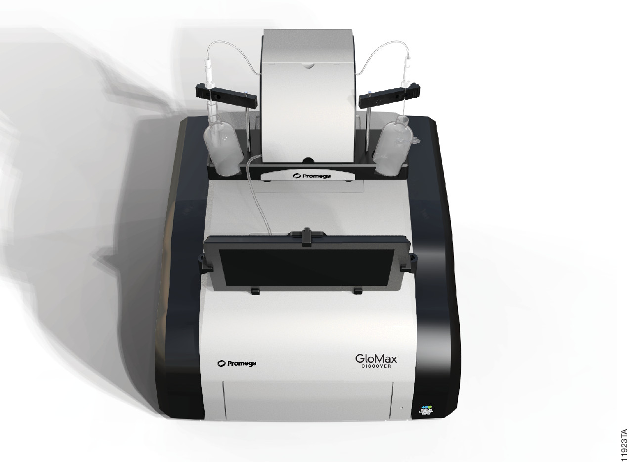
Luciferase assays are useful tools for studying a wide range of biological questions. They can be performed easily by adding a reagent that provides components necessary to generate a luminescent signal directly to cells or a cell lysate. However, once this reagent has been added, how long you wait to measure the signal becomes a key consideration in generating consistent data. Dependent on which luciferase assay you use, you may need a luminometer that can use injectors to deliver the assay reagents. The reason for this is simple, but can be confusing to new users.
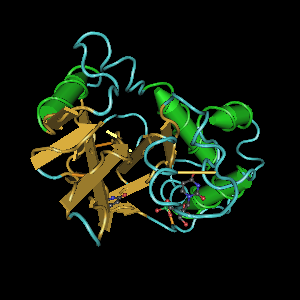
Proteomics, the analysis of the entire protein content of a living system, has become a vital part of life science research, and mass spectrometry (MS) is the method for analyzing proteins. MS analysis of protein content allows researchers to identify proteins, sequence them and determine the nature of post translational modifications.
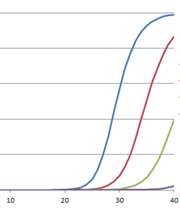
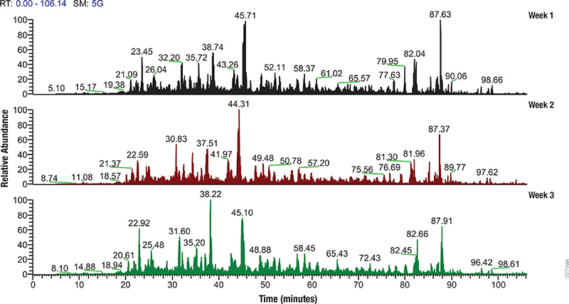
LC/MS performance monitoring. Each run used 1μg of human predigested protein extract injected into the instrument (Waters NanoAquity HPLC System interfaced to a Thermo Fisher Q Exactive™ Hybrid Quadrupole-Orbitrap Mass Spectrometer). Peptides were resolved with a 2-hour gradient. Weekly monitoring with the human extract ensured consistent analytical performance of the instrument.
Mass spectrometry allows characterization of molecules by converting them to ions so that they can be manipulated in electrical and magnetic fields. Basically a small sample (analyte) is ionized, usually to cations by loss of an electron. After ionization, the charged particles (ions) are separated by mass and charge; the separated particles are measured and data displayed as a mass spectrum. The mass spectrum is typically presented as a bar graph where each peak represents a single charged particle having a specific mass-to-charge (m/z) ratio. The height of the peak represents the relative abundance of the particle. The number and relative abundance of the ions reveal how different parts of the molecule relate to each other.
For the study of large, organic macromolecules, matrix associated laser desorption/ionization (MALDI) or tandem mass spec/collision induced dissociation (MS/MS) techniques are often used to generate the charged particles from the analyte. MS analysis brings sensitivity and specificity to proteome analysis. The technique has excellent resolution and is able to distinguish one ion from another, even when their m/z ratios are similar. Macromolecules are present in extremely different concentrations in the cells, and MS analysis can detect biomolecules across five logs of concentration.
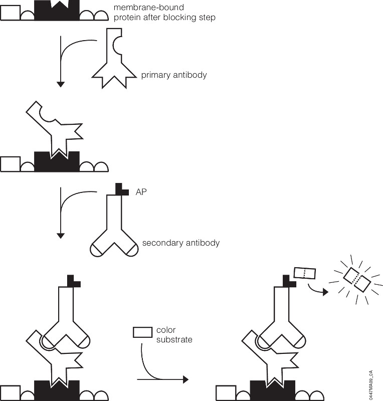
You’ve probably been there. You’ve got a new antibody or you’re testing out one you’ve made yourself. After weeks or months of work, your antibody is going to help move your research project forward. As you excitedly head to the dark room to develop your film, your mood is crushed when you see…bands, more bands, and smears. Alas, science has played one more cruel joke on you as you experience what so many of your fellow scientists have before. Despite such a dismal beginning, you often can still get good western blots by changing steps in your protocol.
Several steps in the western blot protocol can be optimized.
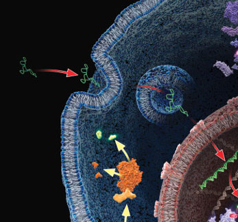
In vitro translation of proteins through cell-free expression systems using rabbit reticulocytes, E. coli S30, or wheat germ extracts can be invaluable in studying protein function. If you only need a small amount (100s of nanograms), it’s also faster and easier than synthesizing vast quantities in bacterial or mammalian cells (~ 90 minutes for cell-free vs. long growth times and extraction steps after an initial optimization for protein synthesized in larger scale). There are many systems out there, and knowing which to use can sometimes be difficult. Many kits include components that combine transcription and translation in one-step, eliminating the need to provide your own RNA. But when you want to make your own RNA templates to add to lysates, then there are additional concerns.
A protein chain being produced from a ribosome.
Many people don’t want to work with RNA since the common lab lore suggests it’s a finicky molecule, and for good reason. Extracting it requires the utmost care in technique and elimination of nucleases. Failing to do so results in degradation of the molecule, and so with it your experiments (see our recent blog by Terri Sundquist on tips for isolating RNA with ease). Preparing RNA for cell-free expression is subject to the same concerns as extracted RNA, but with the proper care is not that much more of a challenge than using a DNA template.
The first step for using cell-free expression systems with RNA templates is to make the RNA. Here are some tips that will ensure success.
This review is a guest blog by Amy Landreman, Product Specialist in Cellular Analysis at Promega Corporation.
Lentiviral vectors (LVV) have become a valuable research tool for delivering genetic content into a wide range of cell types. Commonly derived from the HIV-1 genome, LVV have the advantage of being able to infect both dividing and non-dividing cells. They can be particularly valuable for introducing genetic material into cell lines that are difficult to transfect using other methods and are also being used in gene therapy applications.
Unlike other gene delivery tools, transducing mammalian cells with LVV requires significant upfront effort since the LVV particles carrying the desired genetic content first need to be created. In general this involves co-transfecting a packaging cell line, such as HEK293T, with a set of three to four separate plasmids that encode the protein content required to generate the LVV particles: the transfer plasmid, which contains the transgene of interest, a packaging plasmid, and an envelope plasmid. After co-transfection, the packaging cell line is allowed to incubate for a couple of days during which time the LVV particles are produced and accumulating in the culture supernatant. The supernatant containing the recombinant LVV is then harvested and, following several concentration steps, the LVV particles are ready to be used for introducing the desired genetic content into the mammalian target cells. Continue reading “Get More Out of Your Lentiviral Production”
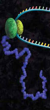
Ribbon diagram of RNA’s biggest threat: a ribonuclease
Back in graduate school, I purified a lot of RNA, and after a while, I became fairly successful at it. My yields were good, and the RNA was intact. However, many of my early attempts at RNA isolation yielded degraded RNA that did not work well in many downstream applications. In my case, successfully isolating high-quality RNA required practice. During my trials and tribulations, I learned a lot of tricks and tips about how to obtain high-quality RNA. Here I share some of these tricks to help you speed through that “practice makes perfect” phase so that you can isolate RNA like a pro.
The polymerase chain reaction (PCR) has revolutionized modern biology as a quick and easy way to generate amazing amounts of genomic data. However, when PCR doesn’t work, it can be frustrating. At these times, PCR and reverse transcription PCR (RT-PCR) inhibitors seem to be everywhere: They lie dormant in your starting material and can co-purify with the template of interest, and they can be introduced during sample handling or reaction setup. The effects of these inhibitors can range from partial inhibition and underestimation of the target nucleic acid amount to complete amplification failure. What is a scientist to do?
HEK293 cells stably expressing HaloTag®-ECS fusion protein labeled with HaloTag® Alexa Fluor® 488 Ligand and then imaged.
G Protein Coupled Receptors represent one of the largest classes of cell surface receptors and one of the most important classes for drug targets. Fifty of the top 200 drugs target GPCRs. GPCRs respond to various stimuli like light, odors, hormones, neurotransmitters and others. They cover virtually all therapeutic areas. When a particular GPCR is implicated in a disease, researchers screen the GPCR and its signaling pathways, the hope being that promising therapeutic targets might be identified. Major G-protein families signal via secondary messengers like cAMP, which in turn activate a range of effector systems to change cell behavior and/or gene transcription. There are various approaches and methods to study GPCRs and measure the increase or decrease of intracellular cAMP. However, the fastest and the most sensitive among all methods is a plate based cAMP-Glo™ Assay.
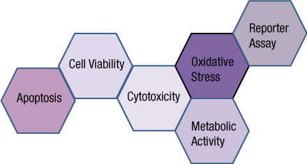
You often need several pieces of information to really understand what is happening within a cell or population of cells. If your cells are not proliferating, are they dying? Or, are you seeing cytostasis? If they are dying, what is the mechanism? Is it apoptosis or necrosis? If you are seeing apoptosis, what is the pathway: intrinsic or extrinsic?
If you are measuring expression of a reporter gene and you see a decrease in expression, is that decrease due to transfection inefficiencies, cytotoxicity, or true down regulation of your reporter gene?
To investigate these multiple parameters, you can run assays in parallel, but that requires more sample, and sample isn’t always abundant.
Multiplexing assays allows you to obtain information about multiple parameters or events (e.g., reporter gene expression and cell viability; caspase-3 activity and cell viability) from a single sample. Multiplexing saves sample, saves time and gives you a more complete picture of the biology that is happening with your experimental sample.
What information do you need about your cells to complete the picture?
Multiplexing assay reagents to measure biomarkers in the same sample has often been considered an application only accomplished with antibodies or dyes and sophisticated detection instrumentation. However, Promega has developed microwell plate based assays for cells in culture that allow multiplexed detection of biomarkers in the same sample well using standard multimode multiwell plate readers. Continue reading “Piecing the Puzzle Together: Using Multiple Assays to Better Understand What Is Happening with Your Cells”
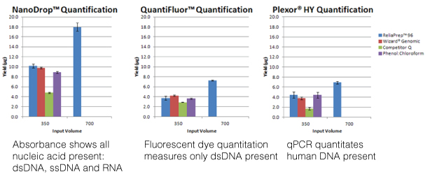
For most molecular biology applications, knowing the amount of nucleic acid present in your purified sample is important. However, one quantitation method might serve better than another, depending on your situation, or you may need to weigh the benefits of a second method to assess the information from the first. Our webinar “To NanoDrop® or Not to NanoDrop®: Choosing the Most Appropriate Method for Nucleic Acid Quantitation” given by Doug Wieczorek, one of our Applications Scientists, discussed three methods for quantitating nucleic acid and outlined their strengths and weaknesses.
XWe use cookies and similar technologies to make our website work, run analytics, improve our website, and show you personalized content and advertising. Some of these cookies are essential for our website to work. For others, we won’t set them unless you accept them. To learn more about our approach to Privacy we invite you to Read More
By clicking “Accept All”, you consent to the use of ALL the cookies. However you may visit Cookie Settings to provide a controlled consent.
We use cookies and similar technologies to make our website work, run analytics, improve our website, and show you personalized content and advertising. Some of these cookies are essential for our website to work. For others, we won’t set them unless you accept them. To find out more about cookies and how to manage cookies, read our Cookie Policy.
If you are located in the EEA, the United Kingdom, or Switzerland, you can change your settings at any time by clicking Manage Cookie Consent in the footer of our website.
Necessary cookies are absolutely essential for the website to function properly. These cookies ensure basic functionalities and security features of the website, anonymously.
Cookie
Duration
Description
cookielawinfo-checbox-analytics
11 months
This cookie is set by GDPR Cookie Consent plugin. The cookie is used to store the user consent for the cookies in the category "Analytics".
cookielawinfo-checbox-functional
11 months
The cookie is set by GDPR cookie consent to record the user consent for the cookies in the category "Functional".
cookielawinfo-checbox-others
11 months
This cookie is set by GDPR Cookie Consent plugin. The cookie is used to store the user consent for the cookies in the category "Other.
cookielawinfo-checkbox-advertisement
1 year
The cookie is set by GDPR cookie consent to record the user consent for the cookies in the category "Advertisement".
cookielawinfo-checkbox-necessary
11 months
This cookie is set by GDPR Cookie Consent plugin. The cookies is used to store the user consent for the cookies in the category "Necessary".
cookielawinfo-checkbox-performance
11 months
This cookie is set by GDPR Cookie Consent plugin. The cookie is used to store the user consent for the cookies in the category "Performance".
gdpr_status
6 months 2 days
This cookie is set by the provider Media.net. This cookie is used to check the status whether the user has accepted the cookie consent box. It also helps in not showing the cookie consent box upon re-entry to the website.
lang
This cookie is used to store the language preferences of a user to serve up content in that stored language the next time user visit the website.
viewed_cookie_policy
11 months
The cookie is set by the GDPR Cookie Consent plugin and is used to store whether or not user has consented to the use of cookies. It does not store any personal data.
Analytical cookies are used to understand how visitors interact with the website. These cookies help provide information on metrics the number of visitors, bounce rate, traffic source, etc.
Cookie
Duration
Description
SC_ANALYTICS_GLOBAL_COOKIE
10 years
This cookie is associated with Sitecore content and personalization. This cookie is used to identify the repeat visit from a single user. Sitecore will send a persistent session cookie to the web client.
vuid
2 years
This domain of this cookie is owned by Vimeo. This cookie is used by vimeo to collect tracking information. It sets a unique ID to embed videos to the website.
WMF-Last-Access
1 month 18 hours 24 minutes
This cookie is used to calculate unique devices accessing the website.
_ga
2 years
This cookie is installed by Google Analytics. The cookie is used to calculate visitor, session, campaign data and keep track of site usage for the site's analytics report. The cookies store information anonymously and assign a randomly generated number to identify unique visitors.
_gid
1 day
This cookie is installed by Google Analytics. The cookie is used to store information of how visitors use a website and helps in creating an analytics report of how the website is doing. The data collected including the number visitors, the source where they have come from, and the pages visted in an anonymous form.
Advertisement cookies are used to provide visitors with relevant ads and marketing campaigns. These cookies track visitors across websites and collect information to provide customized ads.
Cookie
Duration
Description
IDE
1 year 24 days
Used by Google DoubleClick and stores information about how the user uses the website and any other advertisement before visiting the website. This is used to present users with ads that are relevant to them according to the user profile.
test_cookie
15 minutes
This cookie is set by doubleclick.net. The purpose of the cookie is to determine if the user's browser supports cookies.
VISITOR_INFO1_LIVE
5 months 27 days
This cookie is set by Youtube. Used to track the information of the embedded YouTube videos on a website.
Performance cookies are used to understand and analyze the key performance indexes of the website which helps in delivering a better user experience for the visitors.
Cookie
Duration
Description
YSC
session
This cookies is set by Youtube and is used to track the views of embedded videos.
_gat_UA-62336821-1
1 minute
This is a pattern type cookie set by Google Analytics, where the pattern element on the name contains the unique identity number of the account or website it relates to. It appears to be a variation of the _gat cookie which is used to limit the amount of data recorded by Google on high traffic volume websites.
 This review is a guest blog by Amy Landreman, Product Specialist in Cellular Analysis at Promega Corporation.