What do Exosomes have to do with Cancer Research?

microRNA that is inside exosomes

Discovered in 1983 and initially dismissed as ‘cellular dust,’ exosomes have since emerged as pivotal players in biomedical research due to their roles in intercellular communication, potential as drug delivery vectors and as biomarkers for various diseases. These small extracellular vesicles, measuring 30–150nm, are crucial for transferring proteins, lipids, and nucleic acids — including microRNA (miRNA), mRNA, and non-coding RNA– between cells (1). miRNAs are particularly critical as they regulate gene expression and offer insights into the cellular mechanisms underlying diseases like cancer, enhancing the value of exosomes in cancer research.

Beyond exosomes importance in understanding intracellular communication and organ cross-talk, exosomes can also alter the functions of recipient cells based on their cargo. This capability makes them extremely valuable in providing insights into alterations in cellular communication, tumor microenvironments, metastasis and immune evasion.

Continue reading “What do Exosomes have to do with Cancer Research?”
Will Artificial Intelligence (AI) Transform the Future of Life Science Research?

Artificial intelligence (AI) is not a new technological development. The idea of intelligent machines has been popular for several centuries. The term “artificial intelligence” was coined by John McCarthy for a workshop at Dartmouth College in 1955 (1), and this workshop is considered the birthplace of AI research. Modern AI owes much of its existence to an earlier paper by Alan Turing (2), in which he proposed the famous Turing Test to determine whether a machine could exhibit intelligent behavior equivalent to—or indistinguishable from—that of a human.

The explosive growth in all things AI over the past few years has evoked strong reactions from the general public. At one end of the spectrum, some people fear AI and refuse to use it—even though they may have unwittingly been using a form of AI in their work for years. At the other extreme, advocates embrace all aspects of AI, regardless of potential ethical implications. Finding a middle ground is not always easy, but it’s the best path forward to take advantage of the improvements in efficiency that AI can bring, while still being cautious about widespread adoption. It’s worth noting that AI is a broad, general term that covers a wide range of technologies (see sidebar).

AI personified looking at a dna double helix against an abstract cosmic background
Image generated with Adobe Firefly v.2.

For life science researchers, AI has the potential to address many common challenges; a previous post on this blog discussed how AI can help develop a research proposal. AI can help with everyday tasks like literature searches, lab notebook management, and data analysis. It is already making strides on a larger scale in applications for lab automation, drug discovery and personalized medicine (reviewed in 3–5). Significant medical breakthroughs have resulted from AI-powered research, such as the discovery of novel antibiotic classes (6) and assessment of atherosclerotic plaques (7). A few examples of AI-driven tools and platforms covering various aspects of life science research are listed here.

Continue reading “Will Artificial Intelligence (AI) Transform the Future of Life Science Research?”

Monochromator vs Filter-Based Plate Reader: Which is Better?

When it comes to purchasing a microplate reader for fluorescence detection, the most common question is whether to choose a monochromator-based reader or filter-based reader. In this blog, we’ll discuss how both types of plate readers work and factors to consider when determining the best plate reader for your need.

How do monochromator-based plate readers work?

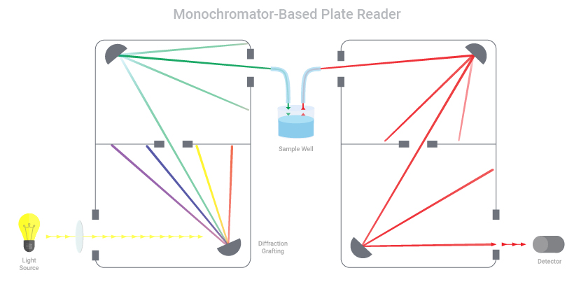
Monochromators work by taking a light source and splitting the light to focus a particular wavelength on the sample. During excitation, the light passes through a narrow slit, directed by a series of mirrors and diffraction grating and then passes through a second narrow slit prior to reaching the sample. This ensures the desired wavelength is selected to excite the fluorophore. Once the fluorophore is excited, it emits light at a different, longer wavelength. This emission light is captured by another series of mirrors, grating and slits to limit the emission to a desired wavelength, which then enters a detector for signal readout.

Monochromator-based plate reader
Continue reading “Monochromator vs Filter-Based Plate Reader: Which is Better?”

Just What Is an RLU (Relative Light Unit)?

This post was contributed by guest blogger, Scott Messenger, Technical Support Scientist 2 at Promega Corporation.

It’s always an exciting time in the lab when you find a new assay to answer an important research question. Once you get your hands on the assay, it is always good to confirm it will work for your experimental setup. Repeating the control experiment shown in the technical manual is a great way to test the assay in your hands.

After running that first experiment of your assay, it looks pretty good. The trends of control and treatment are consistent. Time to get on with the experiments…but wait—the RLUs (Relative Light Units) are two orders of magnitude lower than the example data! I can’t show this data to my colleagues; it doesn’t match. What did I do wrong?

Relative Light Units, Measuring, Luminescence

This is a concern that we in Technical Services hear frequently. The concern is real, and I had this same thought when doing some of my first experiments using luminescence. When a question like this comes in, a Technical Service Scientist will make sure the experiment was performed as we described, and in most cases it is. We then start talking about RLUs (Relative Light Units).

Continue reading “Just What Is an RLU (Relative Light Unit)?”

Why You Don’t Need to Select a Wavelength for a Luciferase Assay

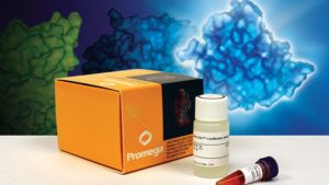
Promega kit depicted; test involves wavelength for a luciferase assay.

It’s a question I’m asked probably once a week. “What wavelength do I select on my luminometer when performing a luciferase assay?” The question is a good and not altogether unexpected one, especially for those new to bioluminescent assays. The answer is that in most cases, you don’t and in fact shouldn’t select a wavelength (the exception to this rule is if you’re measuring light emitted in two simultaneous luciferase reactions). To understand why requires a bit of an explanation of absorbance, fluorescence, and luminescence assays, and the differences among them.

Absorbance, fluorescence, and luminescence assays are all means to quantify something of interest, be that a genetic reporter, cell viability, cytotoxicity, apoptosis, or other markers. In principle, they are all similar. For example, a genetic reporter assay is an indicator of gene expression. The promoter of a gene of interest can be cloned upstream of a reporter such as β-galactosidase, GFP, or firefly luciferase. The amount of each of these reporters that is transcribed into mRNA and translated into protein by the cell is indicative of the endogenous expression of the gene of interest.

Continue reading “Why You Don’t Need to Select a Wavelength for a Luciferase Assay”

Eight Considerations for Getting the Best Data from Your Luminescent Assays

The stage is set. You’ve spent days setting up this experiment. Your bench is spotless. All the materials you need to finally collect data are laid neatly before you. You fetch your cells from the incubator, add your detection reagents, and carefully slide the assay plate into the luminometer. It whirs and buzzes, and data begin to appear on the computer screen. But wait!

Bad data
These data are garbage!

Don’t let this dramatic person be you. Here are 8 tips from us on things to watch out for before you start your next luminescent assay. Make sure you’ll be getting good data before wasting precious sample!

Continue reading “Eight Considerations for Getting the Best Data from Your Luminescent Assays”

Three Factors That Can Hurt Your Assay Results

4621CA

Each luminescent assay plate represents precious time, effort and resources. Did you know that there are three things about your detection instrument that can impact how much useful information you get from each plate?  Instruments with poor sensitivity may cause you to miss low-level samples that could be the “hit” you are looking for.  Instruments with a narrow detection range limit the accuracy or reproducibility you needed to repeat your work.  Finally, instruments that let the signal from bright wells spill into adjacent wells allow crosstalk to occur and skew experimental results, costing you time and leading to failed or repeated experiments.

Continue reading “Three Factors That Can Hurt Your Assay Results”

To inject or not inject?

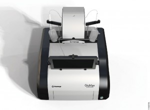
GloMax® Discover Multimode Reader with injectors.

Luciferase assays are useful tools for studying a wide range of biological questions. They can be performed easily by adding a reagent that provides components necessary to generate a luminescent signal directly to cells or a cell lysate. However, once this reagent has been added, how long you wait to measure the signal becomes a key consideration in generating consistent data. Dependent on which luciferase assay you use, you may need a luminometer that can use injectors to deliver the assay reagents. The reason for this is simple, but can be confusing to new users.

Let’s start by discussing two types of luciferase assays: “flash” vs. “glow”. Continue reading “To inject or not inject?”