Discovering the Complexity of the Human Proteome

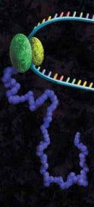
Transcription TranslationI should preface this blog by stating that I am a nucleic acids gal. My years in the lab were spent with tubes of DNA and RNA. In fact my one and only tentative foray into the field of protein resulted in a Western Blot so ugly that those who witnessed it have been sworn to secrecy. Given all of this, the mapping of the human proteome might seem like an odd topic for me to write about. Except that it isn’t really, because the sequencing of the proteome offers answers to some of the questions that the sequencing of the genome didn’t.

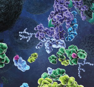
First, let’s start with what a proteome is: A proteome is all the proteins expressed at a certain time point. It can be as limited as the proteome of a single cell or as all encompassing as the proteome of an entire genome. However, unlike the genome, which is genetic information encoded in an organism’s DNA or RNA, the makeup of a proteome can vary dramatically as a result of expression patterns, alternative splicing events and post-translational modifications.

The genome is a constant, what you see today is what will still be there tomorrow. The proteome, on the other hand, is a constantly changing landscape. Up regulation or down regulation of a gene can mean more or less protein is present. Alternative splicing and post-translational modifications can result in fundamental changes to the protein itself.

In other words, if the genome is a beautiful, pristine Ansel Adams print, then the proteome is that same scene as interpreted by Andy Warhol—in Technicolor and 3D. Continue reading “Discovering the Complexity of the Human Proteome”

Purifying HIS-Tagged Proteins from Insect and Mammalian Cells

MSextractcroppedMany different polypeptide fusion partners or affinity tags have been developed to facilitate purification of target proteins. The most commonly used tag for the purification and detection of recombinant expressed proteins is the His tag. Cloning vectors designed to generate His-tagged proteins contain 5–10 histidine residues at either the C- or N terminus of the expressed protein. The His tag adds only 0.84kDa to the mass of the protein and is nonimmunogenic. Also, because the tertiary structure of the tag is not important for purification, His-tagged proteins can be purified using native or denaturing conditions. The affinity of histidine residues for immobilized nickel allows selective purification of His-tagged proteins. The MagneHis™ Ni-Particles can bind up to 1mg of His-tagged protein per milliliter of particles providing a fast, efficient method for purifying His-tagged proteins with high yield and low background in a highly scalable format.
Bacterial expression of recombinant His-tagged proteins is a common technique. However, use of other systems, such as Sf9 insect cells,or HeLa or CHO mammalian cells for expression of recombinant proteins either intracellularly or secreted into the culture medium is increasing. These eukaryotic expression systems may allow more natural processing and modification of recombinant His-tagged proteins.
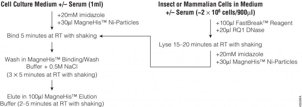
The following article:  illustrates the use of FastBreak™ Cell Lysis Reagent and the MagneHis™ Protein Purification System with insect and mammalian cell lysates. Proteins are purified from culture medium in the presence or absence of serum with only minior modifications to the standard protocol for bacterial cultures are required for purification from these diverse sources.
4550MA-(1)


Monitoring Mass Spec Instrument Performance and Sample Preparation

Proteomics, the analysis of the entire protein content of a living system, has become a vital part of life science research, and mass spectrometry (MS) is the method for analyzing proteins.  MS analysis of protein content allows researchers to identify proteins, sequence them and determine the nature of post translational modifications.

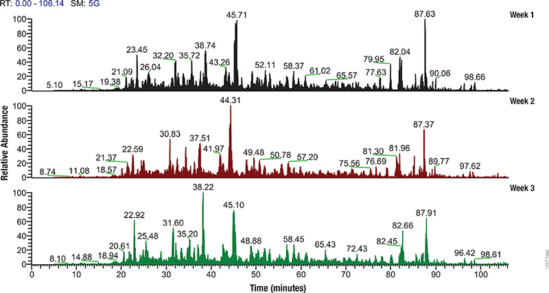
LC/MS performance monitoring. Each run used 1μg of human predigested protein extract injected into the instrument (Waters NanoAquity HPLC System interfaced to a Thermo Fisher Q Exactive™ Hybrid Quadrupole-Orbitrap Mass Spectrometer). Peptides were resolved with a 2-hour gradient. Weekly monitoring with the human extract ensured consistent analytical performance of the instrument.

Mass spectrometry allows characterization of molecules by converting them to ions so that they can be manipulated in electrical and magnetic fields. Basically a small sample (analyte) is ionized, usually to cations by loss of an electron. After ionization, the charged particles (ions) are separated by mass and charge;  the separated particles are measured and data displayed as a mass spectrum. The mass spectrum is typically presented as a bar graph where each peak represents a single charged particle having a specific mass-to-charge (m/z) ratio. The height of the peak represents the relative abundance of the particle. The number and relative abundance of the ions reveal how different parts of the molecule relate to each other.

For the study of large, organic macromolecules, matrix associated laser desorption/ionization (MALDI) or tandem mass spec/collision induced dissociation (MS/MS) techniques are often used to generate the charged particles from the analyte. MS analysis brings sensitivity and specificity to proteome analysis. The technique has excellent resolution and is able to distinguish one ion from another, even when their m/z ratios are similar. Macromolecules are present in extremely different concentrations in the cells, and MS analysis can detect biomolecules across five logs of concentration.

Continue reading “Monitoring Mass Spec Instrument Performance and Sample Preparation”

Shedding Light on Protein:Protein Interactions with NanoBRET™ Technique

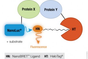
NanoBRET™ TechnologyIf you are trying to investigate protein:protein interactions inside cells, you know how important physiologically relevant results are. If you overload your cells with fusion constructs, your protein interactions may not actually reflect what is going on in the cell, and if your BRET energy donor and acceptor do not have sufficiently separated spectra, you can pick up a fair amount of noise in your experiment. Using the new superbright NanoLuc® Luciferase, and the HaloTag® Technology, we have developed a sensitive BRET system to help you take a better look specific protein interactions that interest you. Promega research scientist, Danette Daniels, describes the system in the Chalk Talk below:

Optimize Your Western Blot

0447MA09_0A
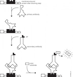
Western Blot Detection.

You’ve probably been there. You’ve got a new antibody or you’re testing out one you’ve made yourself. After weeks or months of work, your antibody is going to help move your research project forward. As you excitedly head to the dark room to develop your film, your mood is crushed when you see…bands, more bands, and smears. Alas, science has played one more cruel joke on you as you experience what so many of your fellow scientists have before. Despite such a dismal beginning, you often can still get good western blots by changing steps in your protocol.

Several steps in the western blot protocol can be optimized.

Continue reading “Optimize Your Western Blot”

Mass Spec-Compatible Proteome Reference Material

MSextractcropped

The complexity of biological samples places high demand on mass spec analytical capability. Adequate monitoring of instrument performance for proteomics studies requires equally complex reference material such as whole-cell extracts. However, whole-cell extracts available commercially are developed for general research (e.g., enzymatic or Western blot analysis) and contain detergents and salts that interfere with reverse phase liquid chromatography and mass spectrometry. Even after clean up, the extracts need to be digested, requiring time, labor and experience to generate samples for use in mass spectrometry. To address the need for complex protein material, we have developed whole-cell protein extracts from yeast and human cells. The yeast extract offers the convenience of a relatively small and well annotated proteome, whereas the human extract provides a complex proteome with large dynamic range. The human extract also serves as reference material for studies targeting the human proteome.

The extracts are free of compounds that interfere with reverse phase liquid chromatography-mass spectrometry (LC-MS), and have been reduced with DTT and alkylated with iodoacetamide then digested with Trypsin/Lys-C Mix and lyophilized. These digested extracts (tryptic peptides) can be readily reconstituted in trifluoroacetic acid (TFA) or formic acid and injected into an instrument. The same human and yeast whole-cell extracts also are provided in an intact (undigested) form for users who would like to develop an independent method for preparing protein mass spectrometry samples. For convenience, the intact extracts are provided as a frozen solution.

Consistent extract protein composition is ensured by tight control over cell culture conditions and manufacturing process. Lot-to-lot consistency of extracts is monitored by various protein and peptide qualitative and quantitation methods, including LC-MS. (Quality control results are provided upon request.) Our manufacturing process assures compatibility with reverse phase liquid chromatography and mass spectrometry, minimal nonspecific protein fragmentation, nonbiological post-translational modifi cations and,for digested extracts, minimal undigested peptides. The extracts are optimized for a high number of peptide and protein identifications in mass spectrometry analysis.

His-Tagged Fusion Proteins: Application Update

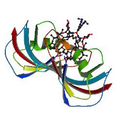
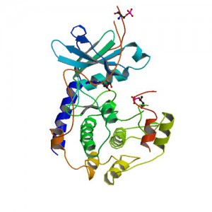
Crystal structure of Polyhistidine tagged recombinant catalytic subunit of cAMP-dependent protein kinase. Credit: StructureID=1fmo; DOI=http://dx.doi.org/10.2210/pdb1fmo/pdb;

Researchers often need to purify a single protein for further study. One method for isolating a specific protein is the use of affinity tags. Affinity purification tags can be fused to any recombinant protein of interest, allowing fast and easy purification following a procedure that is based on the affinity properties of the tag.

The most commonly used tag to purify and detect recombinant expressed proteins is the polyhistidine tag. Protein purification using polyhistidine tags relies on the affinity of histidine residues for immobilized metal such as nickel, which allows selective protein purification. The metal is immobilized to a support through complex formation with a chelate that is covalently attached to the support.

Polyhistidine tags offer several advantages for protein purification. The small size of the polyhistidine tag renders it less immunogenic than other larger tags. Therefore, the tag usually does not need to be removed for downstream applications following purification.

A large number of commercial expression vectors that contain polyhistidine are available. The polyhistidine tag may be placed on either the N- or C-terminus of the protein of interest.

And finally, the interaction of the polyhistidine tag with the metal does not depend on the tertiary structure of the tag, making it possible to purify otherwise insoluble proteins using denaturing conditions. The resulting purified protein can be used for a variety of applications.

The following references illustrate examples of some of the most common post purification applications with fusion proteins containing a polyhistidine tag:

Enzymatic assays

  1. Negi, V-S. et al. (2014) A carbon nitrogen Lyase from Leucaena leucocephala catalyzes the first Step of mimosine degradationPlant Physiol. 164,  922–34.
  2. Yu, S. et al. (2013) Syk Inhibits the activity of protein kinase A by phosphorylation tyrosine of the catalytic subunitJ. Biol. Chem. 288, 10870-81.
  3. Rusconi, B. et al. (2013) Discovery of catalases in members of the Chiamydiales order. J. Bact. 195, 3543–51.

Structural analysis

  1. Araiso, Y. et al. (2014) Crystal structure of Saccharomyces cerevisiae mitochondrial GatFAB a novel subunit assembly in tRNA –dependent amidotransferases. Nucl.Acids. Res.(available only online).
  2. Someya, T. et al. (2012) Crystal Structure of Hfq from Bacillus subtilis in complex with SELEX-dervived RNA aptamer: insight into RNA-binding properties of bacterial Hfq. Nucl. Acid Res. 40, 1856-67.

Protein pulldowns

  1. Yun, S-C. et al. (2010) Pmr a Histone-like protein H1 (H-NS) family protein encoded by the IncP-7 plasmid pCAR1, is a key global regulator that alters host functionJ.Bact. 192, 4720–31.
  2. Haim, H. et al. (2010)  Cytokeratin 8 interacts with clumping factor B: a new possible virulence factor target. Microbiology 156, 3710-21.

Additional Resources
His-tagged Protein Purification Systems

CheckMate™ Mammalian Two-Hybrid System: Application Update

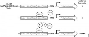
Assay principle for CheckMate™ Mammalian Two-Hybrid System.
Assay principle.

In the CheckMate™ Mammalian Two-Hybrid System, the pBIND Vector contains the yeast GAL4 DNA-binding domain upstream of a multiple cloning region, and the pACT Vector contains the herpes simplex virus VP16 activation domain upstream of a multiple cloning region. The two genes encoding the two potentially interactive proteins of interest are cloned into pBIND and pACT Vectors to generate fusion proteins with the DNA-binding domain of GAL4 and the activation domain of VP16, respectively. The pG5luc Vector contains five GAL4 binding sites upstream of a minimal TATA box, which in turn, is upstream of the firefly luciferase gene (luc+). The pGAL4 and pVP16 fusion constructs are transfected along with pG5luc Vector into mammalian cells. Interaction between the two test proteins, as GAL4 and VP16 fusion constructs, results in an increase in firefly luciferase expression over the negative controls. Traditionally mammalian two hybrid analysis was used to confirm initial data obtained from yeast two hybrid experiments.

Due to enhanced bioinformatics information and the development of improved co-immunoprecipiation/pulldown procedures/technology, there is a growing trend to use only mammalian cells to characterize protein:protein interactions. The following references illustrate the use of the CheckMate™ system to complement  other techniques to characterize protein;protein interactions using only mammalian cells .

Bagchi, P. et al. (2013) Molecular Mechanism behind Rotavirus Nsp-1 Mediated PI3 Kinase Activation: Interaction between NSP1 and the p85Subunit of PI3 kinase. J. Vir. 87, 2358-62.

Greninger, A. et al. (2013) ACBD3 Interaction with TBC1 domain 22 protein is differentially affected by Enteroviral and Kobuviral 3A protein binding.  mBio 4, 00098-13.

Patki, M. et al. ( 2013) The ETS Domain Transcription Factor ELK1 Direct a critical component of growth signalling by Androgen Receptor in prostrate cancer cells. J.Biol. Chem. 288 11047-65

Konig, H-G. (2012) Fibroblast growth factor homologous factor 1 interact with NEMO to regulate NF-kappaB signaling in neurons J. Cell Sci. 125, 6058-70

Lost in Translation? Tips for Preparing RNA for in vitro Translation Experiments

In vitro translation of proteins through cell-free expression systems using rabbit reticulocytes, E. coli S30, or wheat germ extracts can be invaluable in studying protein function.  If you only need a small amount (100s of nanograms), it’s also faster and easier than synthesizing vast quantities in bacterial or mammalian cells (~ 90 minutes for cell-free vs. long growth times and extraction steps after an initial optimization for protein synthesized in larger scale).  There are many systems out there, and knowing which to use can sometimes be difficult.  Many kits include components that combine transcription and translation in one-step, eliminating the need to provide your own RNA.  But when you want to make your own RNA templates to add to lysates, then there are additional concerns.

artists concept of in vitro translation
A protein chain being produced from a ribosome.

Many people don’t want to work with RNA since the common lab lore suggests it’s a finicky molecule, and for good reason.  Extracting it requires the utmost care in technique and elimination of nucleases.  Failing to do so results in degradation of the molecule, and so with it your experiments (see our recent blog by Terri Sundquist on tips for isolating RNA with ease).  Preparing RNA for cell-free expression is subject to the same concerns as extracted RNA, but with the proper care is not that much more of a challenge than using a DNA template.

The first step for using cell-free expression systems with RNA templates is to make the RNA.  Here are some tips that will ensure success.

Continue reading “Lost in Translation? Tips for Preparing RNA for in vitro Translation Experiments”

Asp-N Protease: Applications Update

TrypsinLysC Page 2

Asp-N, Sequencing Grade, is an endoproteinase that hydrolyzes peptide bonds on the N-terminal side of aspartic and cysteic acid residues: Asp and Cys. Asp-N activity is optimal in the pH range of 4.0–9.0. This sequencing grade enzyme can be used alone or in combination with trypsin or other proteases to produce protein digests for peptide mapping applications or protein identification by peptide mass fingerprinting or MS/MS spectral matching. It is suitable for  in-solution or in-gel digestion reactions.

The following references illustrate the use of Asp-N in recent publications:

Protein sequence coverage

  1. Jakobsson, M et al. (2013)  Identification and characterization of a novel Human Methyltransferase modulating Hsp70 protein function through lysine methylation. J. Biol. Chem. 288, 27752–63.
  2. Carroll, J. et. al. (2013) Post-translational modifications near the quinone binding site of mammalian complex I.  J. Biol. Chem. 288, 24799–08.

Glycoprotein analysis

  1. Siguier, B. et al. (2014) First structural insights into α-L-Arabinofuranosidases from the two GH62 Glycoside hydrolase subfamilies. J. Biol. Chem. 289, 5261–73.
  2. Vakhrushev, S. et al. (2013) Enhanced mass spectrometric mapping of the human GalNAc-type O-glycoproteome with SimpleCells. Mol. Cell. Prot. 12, 932–44.
  3. Berk, J. et al. (2013) . O-Linked β-N- Acetylglucosamine (O-GlcNAc) Regulates emerin binding to autointegration Factor (BAF) in a chromatin and Lamin B-enriched “Niche”.  J. Biol. Chem. 288, 30192–09.

Phosphoprotein analysis

  1. Roux, P. and Thibault, P. (2013) The Coming of Age of phosphoproteomics –from Large Data sets to Inference of protein Functions. Mol. Cell. Prot. 12, 3453–64.

Are you looking for proteases to use in your research?
Explore our portfolio of proteases today.