High-Throughput Screening for Potential Biomarkers Using Cerebrospinal Fluid (CSF)

3240CA02_1A_rename_3Cerebrospinal fluid (CSF) is a bodily fluid present around the brain and in the spinal cord. It acts as a protective cushion against shocks and participates in the immune response in the brain. Analysis of total CSF protein can be used for diagnostic purposes, as, for instance, a sign of a tumor, bleeding, inflammation, or injury. Considering the high value of CSF as a source of potential biomarkers for brain-associated damages and pathologies, the development of robust automated platform for CSF proteomics is of great value.
The scalable automated proteomic pipeline (ASAP2)  was initially developed with the purpose of (i) discovering protein biomarkers in plasma (1). A summary of the ASAP2 process is as follows:As a first step, abundant-protein immuno-affinity depletion is performed with antibody-based columns and LC systems equipped with a refrigerated autosampler and fraction collector. This block is linked to and followed by buffer exchange performed in a 96-well plate format by manual operations that require <1 h to be completed. The rest of the process is fully automated and includes (i) reduction, alkylation, enzymatic digestion.; (ii) tandem mass tag (TMT) labeling and pooling (processing time of ); (iii) RP solid-phase extraction (SPE) purification ; and (iv) strong cation-exchange (SCX) SPE purification.

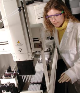
A recent reference (2) validated the use of ASAP2 for sample preparation and proteomic analysis of human CSF samples was performed. CSF samples were first depleted from abundant proteins by multiplexed immuno-affinity. Subsequently, reduction, alkylation, protein digestion (using Trypsin/Lys-C), TMT 6-plex labeling, pooling, and sample cleanup were performed in a 96-well-plate format using a liquid-handling robotic platform. Ninety-six  identical CSF samples were prepared using the highly automated ASAP2 procedure. Proteome coverage consistency, quantitative precision, and individual protein variability, were determined. Results indicated that, ASAP2 is efficient in analyzing large numbers of human CSF samples and would be a valuable tool for biomarker discovery.

References

  1. Dayon, L et al. (2014) Comprehensive and Scalable Highly Automated MS-Based Proteomic Workflow for Clinical Biomarker Discovery in Human Plasma. J of Proteome Res. 13, 3837–45
  2. Galindo, M-N. et al. (2015) Proteomics of Cerebrospinal Fluid: Throughput and Robustness Using a Scalable Automated Analysis Pipeline for Biomarker Discovery. Anan. Chem. 87, 10755–61

Characterizing Unique Protein: DNA Interactions Using Cell-Free Protein Expression

Molecular model of human telomere DNA

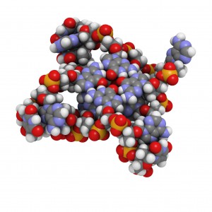
The POT1 protein plays a critical role in telomere protection and telomerase regulation. POT1 binds single-stranded 5′-TTAGGGTTAG-3′ and forms a dimer with the TPP1 protein. Human POT1 contains two Oligonucleotide/Oligosaccharide Binding (OB) fold domains, OB1 and OB2, which make physical contact with the DNA. OB1 recognizes 5′-TTAGGG whereas OB2 binds to the downstream TTAG-3′ (1,2). Several recent studies from other species have shown that some of these proteins are able to recognize a broader variety of DNA ligands than expected (3). A recent reference reexamined the sequence-specificity of the Human POT1 protein (4).
SELEX (Systematic Evolution of Ligands through Exponential Enrichment) was used  to re-examine the DNA-binding specificity of human POT1 (5).

Continue reading “Characterizing Unique Protein: DNA Interactions Using Cell-Free Protein Expression”

Optimizing Antibody Internalization Assays: pHAb Dyes

22255190_pHabAmine_3D_image_050615-final-large

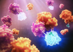
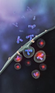
Promega has recently developed a method that allows antibodies to be screened for their internalization properties in a simple, plate-based format. The method uses pH sensor dyes (pHAb dyes), which are not fluorescent at neutral pH but become highly fluorescent at acidic pH. When an antibody conjugated with pHAb dye binds to its antigen on the cancer cell membrane, the antibody-dye-antigen complex is not fluorescent, but upon internalization and trafficking into endosomal and lysosomal vesicles the pH drops, and the dye becomes fluorescent.

To demonstrate the broad utility of the pHAb dye for receptor mediated antibody internalization, two therapeutic antibodies, trastuzumab and cetuximab,which bind to HER2 and EGFR respectively, were selected for a case study (1). Both the antibodies, which are known to internalize were labeled with pHAb dyes using amine or thiol chemistry.

Parameters such as the impact of dye–to-antibody ratio on the antigen–antibody binding, change in fluorescence as a function of pH of free dye and labeled dye, and labeled antibody internalization as a function of pHAb conjugated antibody concentration were evaluated.

The results indicate that pHAb dyes are pH sensitive fluorescent dyes that enable the study of receptor-mediated antibody internalization.Internalization assays can be performed in a plate-based homogeneous format and allow endpoint assays as well as real-time monitoring of internalization. They further show that internalization can be monitored even at a very low amount of antibody which is very important during the early monoclonal antibody development phase when the amount of sample is limited and the antibody concentration in the samples is low. a complimentary approach, they  also showed that a secondary antibody labeled with pHAb dye can be used instead of labeling primary antibodies.

Literature cited

Nath, N. et al. (2016) Homogeneous plate based antibody internalization assay using pH sensor fluorescent dye J.  Immunol. Methods epub ahead of print

Dino Protein: New Methods for Old (Very) Samples

Hadrosaurus skeleton vintage engraving.

Brachylophosaurus was a mid-sized member of the hadrosaurid family of dinosaurs living about 78 million years ago, and is known from several skeletons and bonebed material from the Judith River Formation of Montana and the Oldman Formation of Alberta. Recent fossil evidence indicates structures similar to blood vessels in location and morphology, have been recovered after demineralization of multiple dinosaur cortical bone fragments from multiple specimens, some of which are as old as 80 Ma. These structures were hypothesized to be either endogenous to the bone (i.e., of vascular origin) or the result of biofilm colonizing the empty  network after degradation of original organic components (i.e., bacterial, slime mold or fungal in origin).  Cleland et al. (1) tested the hypothesis that these structures are endogenous and thus retain proteins in common with extant archosaur blood vessels that can be detected with high-resolution mass spectrometry and confirmed by immunofluorescence.

Continue reading “Dino Protein: New Methods for Old (Very) Samples”

Studying Mitochondrial Fission with NanoBiT Complementation Assay

3D depiction of NanoBiT Protein Complementation

Updated February 15, 2021

If you’re interrogating two proteins to understand the conditions under which they interact, a complementation system enables you to tag each protein. Interaction of the tagged proteins facilitates the complementation of the subunits, resulting in a signal. Here we discuss the NanoBiT complementation assay and describe its use to study mitochondrial fission.

Continue reading “Studying Mitochondrial Fission with NanoBiT Complementation Assay”

ProteaseMAX: A Surfactant for the Most Complex Mixtures

Alternate Proteases Cover

Here we provide two examples of “atypical” experiments that take advantage of the properties of the ProteaseMAX™ Surfactant to improve studies involving digestion of complex protein mixtures.

Example 1
Clostridium difficile spores are considered the morphotype of infection, transmission and persistence of C. difficile infections. A recent publication (1) illustrated a novel strategy using three different approaches  to identify proteins of the exosporium layer of C. difficile spores and complements previous proteomic studies on the entire C. difficile spores.

Continue reading “ProteaseMAX: A Surfactant for the Most Complex Mixtures”

NanoBiT™ Assay: Transformational Technology for Studying Protein Interactions Named a Top 10 Innovation of 2015

21416943-wb-cr-uw-nanobit-pm-page-hero
For three out of the last four years, we have been honored to have one of our key technologies named a Top 10 Innovation by The Scientist. This year the innovative NanoBiT™ Assay (NanoLuc® Binary Technology) received the recognition. NanoBiT™ is a structural complementation reporter based on NanoLuc® Luciferase, a small, bright luciferase derived from the deep sea shrimp Oplophorus gracilirostris.

Using plasmids that encode the NanoBiT complementation reporter, you can make fusion proteins to “report” on protein interactions that you are studying. One of the target proteins is fused to the 18kDa subunit; the other to the 11 amino acid subunit. The NanoBiT™ subunits are stable, exhibiting low self-affinity, but produce an ultra-bright signal upon association. So, if your target proteins interact, the two subunits are brought close enough to each other to associate and produce a luminescent signal. The strong signal and low background associated with a luminescent system, and the small size of the complementation reporter, all help the NanoBiT™ assay overcome the limitations associated with traditional methods for studying protein interactions.

The small size reduces the chances of steric interference with protein interactions. The ultra bright signal, means that even interactions among proteins present in very low amounts can be detected and quantified–without over-expressing large quantities of non-native fusion proteins and potentially disrupting the normal cellular environment. And the NanoBiT™ assay can be performed in real time, in live cells.

The NanoBiT™ assay is already being deployed in laboratories to help advance understanding of fundamental cell biology. You can see how one researcher is already taking full advantage of this innovative technology in the video embedded below:

Visit the Promega web site to see more examples more examples how the NanoBiT™ assay can break through the traditional limitations for studying protein interactions in cells.

You can read the Top 10 article in The Scientist here.

IdeZ Protease: A New Tool for the Characterization of Antibodies

12335MB

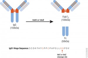
Therapeutic monoclonal antibodies are large, complex molecules that undergo numerous post translational modifications (PTMs).  In-depth characterization of antibody PTMs remains a significant hurdle because their large size (~150 kDa) makes mass spectrometry analysis extremely challenging.

IdeS protease specifically cleaves IgGs into Fab and Fc fragments. This enzyme is highly specific and cleaves human IgG specifically at one site in the lower hinge region.  Because of the exquisite specificity of the enzyme, it produces highly homogeneous Fc and Fab fragments which are then readily analyzed using techniques such as mass spectrometry or HPLC.

Are you looking for proteases to use in your research?
Explore our portfolio of proteases today.

One of the drawbacks of IdeS is that it exhibits poor activity against mouse IgGs. IdeZ Protease is an immunoglobulin-degrading enzyme from Streptococcus equi subspecies zooepidemicus. It is an engineered recombinant protease overexpressed in E. coli. Like IdeS Protease, IdeZ Protease specifically cleaves IgG molecules below the hinge region to yield F(ab′)2 and Fc fragments.  Reduction of the digestion products produces three fragments of ~25kDa that are readily analyzed by LC-MS.

One of the key advantages of the IdeZ Protease is that it has significantly improved activity against mouse IgG2a and IgG3 subclasses compared to IdeS Protease. IdeZ Protease does not cleave mouse IgG1 or IgG2b.

Key technical parameters when digesting mouse IgGs utilizing IdeZ are the following:

• Add 1 unit of IdeZ Protease per 1µg of IgG to be digested.
• IdeZ Protease is most active in buffers at or near neutral pH. The recommended digestion buffer is 50mM sodium phosphate, 150mM NaCl (pH 6.6).
• Mouse IgG2a and IgG3 typically require 2–4 hours at 37°C  for complete digestion.
• IdeZ Protease has a histidine tag for easy removal if so desired.

Interrogating Protein Interactions: An Infographic for NanoBRET™ Assay Design

Yesterday my fellow blogger, Kari, posted a review of the ACS Chemical Biology paper describing a new BRET platform for analyzing protein-protein interactions. If you are interested in studying induction and inhibition of protein interactions in real time, take a look at the infographic below to learn how to develop a NanoBRET™ Assay to monitor your protein of interest.

21421668-IG-CR-NanoBRET-Infographic-Final

Purify and Conjugate Antibodies in a Single Workflow

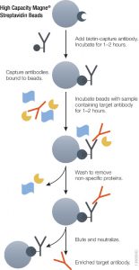
Isoform_Antibodies_LinkedInAntibodies labeled with small molecules such as fluorophore, biotin or drugs play a critical role in various areas of biological research,drug discovery and diagnostics. There are several limitations to current methods for labeling antibodies including the need for purified antibodies at high concentrations and multiple buffer exchange steps.

In a recent publication, a method (on-bead conjugation) is described that addresses these limitations by combining antibody purification and conjugation in a single workflow. This method uses high capacity-magnetic Protein A or Protein G beads to capture antibodies directly from cell media followed by conjugation with small molecules and elution of conjugated antibodies from the beads.

Using a variety of fluorophores the researchers show that the on-bead conjugation method is compatible with both thiol- and amine-based chemistry.

This method enables simple and rapid processing of multiple samples in parallel with high-efficiency antibody recovery. It is further shown that recovered antibodies are functional and compatible with downstream applications.

Literature Cited

Nidhi, N. et al. (2015) On-bead antibody-small molecule conjugation using high-capacity magnetic bead J. Immunol. Methods  http://dx.doi.org/10.1016/j.jim.2015.08.008