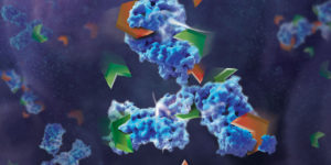
Tetanus Neurotoxin: Potential Mechanism for Drug Delivery

Tetanus neurotoxin (TeNT), produced by Clostridium tetani, is one of the most potent neurotoxins in humans. TeNT causes tetanus, which is characterized by painful muscular contractions and spasms as well as seizure. TeNT is composed of a light chain and a heavy chain (TTH). The toxic properties of TeNT reside in the toxin light chain (L), but like complete TeNT, the TeNT heavy chain (TTH) and the C-terminal domain (TTC) alone can bind and enter into neurons.

Based on these properties, a recent publication (1) considered that TTC could be a promising vehicle to deliver drug cargos to neurons. To explore this possibility, they engineered fusion proteins containing various Tetanus neurotoxin fragments. They chose B-cell leukemia/lymphoma 2 protein (Bcl-2) as a partner protein, because Bcl-2 is one of the most potent anti-apoptotic proteins and has an appropriate size (26kDa) to act as a fusion partner.

They tested these fusion proteins in both cell-based and cell-free protein expression systems to determine whether the purified fusion products retained both anti-apoptotic and neuronal migration properties. One construct (Bcl2-hTTC) exhibited neuronal binding and prevented cell death of neuronal PC12 cells induced by serum and NGF deprivation, as evidenced by the inhibition of cytochrome C release from the mitochondria. For in vivo assays, Bcl2-hTTC was injected into the tongues of mice and was seen to selectively migrate to hypoglossal nuclei mouse brain stems.

  1. Watanbe, Y. et. al. (2018) Tetanus toxin fragments and Bcl-2 fusion proteins : cytoprotection and retrograde axonal migration. BMC Biotechnology 18, 39.

New Recombinant Asp-N Mass Spec Protease: Improved Format and Reduced Price

Asp-N is a endoproteinase hydrolyzes peptide bonds on the N-terminal side of aspartic residues. The native form is isolated from Pseudomonas fragi. The majority of vendors currently provide a commercial product that consists of 2µg of lyophilized material in a flat bottom vial, and sold for $175–200 US. Formatting such a small amount of material in flat bottom vial can lead to inconsistent resuspension of the protease. Inconsistent working concentrations will lead to non-reproducible data. The current high price also prohibits large-scale use.

Are you looking for proteases to use in your research?
Explore our portfolio of proteases today.

The new recombinant Asp-N protease is cloned from Stenotrophomonas maltophilia and expressed in E. coli. Recombinant Asp-N has similar amino acid cleavage specificity as compared to native Asp-N. Digestion of a yeast extract with native and recombinant Asp-N produces very similar results. Providing 10µg lyophilized material in V-shaped vial with a visible cake enables more consistent re-suspension resulting in reproducible data. Due to improved yields the list price is now approximately 40% less when compared to native enzyme.
Learn more about this new recombinant Asp-N protease.

Characterizing Multi-Subunit Protein Complexes Using Cell-Free Expression

artist's concept of a cell membraneMulti-subunit protein complexes control membrane fusion events in eukaryotic cells (1). CORVET and HOPS are two such multi-subunit complexes, both containing the Sec1/Munc18 protein subunit VPS33A (2). Metazoans additionally possess VPS33B, which has considerable sequence similarity to VPS33A but does not integrate into CORVET or HOPS complexes and instead stably interacts with VIPAR. Recent research suggests that VPS33B and VIPAR comprise two subunits of a novel multi-subunit complex analogous in configuration to CORVET and HOPS (3).

In a recent publication (4), Hunter and colleagues, further characterized the VPS33B and VIPAR complex. Using co-immunoprecipitation and proximity-based ligation assay, they identified two novel VPS33B-interacting proteins, VPS53 and CCDC22.

In vitro binding experiments, VPS33B and GST-VIPAR were co-expressed in Escherichia coli and purified by GSH affinity. The VPS33B/GSTVIPAR complex was used as bait in pulldown experiments, with myc-CCDC22 and myc-VPS53 expressed by cell-free in vitro transcription/translation in wheat germ lysate. Myc-CCDC22 was very efficiently pulled down by VPS33B/GST-VIPAR, whereas myc-VPS53 was not .The interaction between VPS53 and the VPS33B-VIPAR complex was either indirect, requires other proteins contribute to the interaction, or requires a post-translational modification not conferred in the plant cell-free expression system (wheat germ). Pull-down experiments with individual subunits or expressing as complexes, was inefficient and did not result in binding to VPS33B/GST-VIPAR.

To further understand how VPS33B-VIPAR may interact with CCDC22, Hunter and colleagues attempted to refine the region of CCDC22 that interacts with VPS33B/GST-VIPAR by generating a series of truncated forms of CCDC22. However, none of five CCDC22 truncations were able to bind to VPS33B/GST-VIPAR. The hypothesis was that truncated forms of CCDC22 are unstable and unable to fold correctly in this assay system.

Additional experiments noted that the protein complex in HEK293T cells which contained VPS33B and VIPAR was considerably smaller than CORVET/HOPS, suggesting that, unlike VPS33A, VPS33B does not assemble into a large stable multi-subunit protein complex.


  1. D’Agostino, M. et. al. (2017) A tethering complex drives the terminal stage of SNARE-dependent membrane fusion. Nature 551, 634–638.
  2. Balderhaar, H. J. K. and Ungermann, C. (2013) CORVET and HOPS tethering complexes – coordinators of endosome and lysosome fusion. J. Cell Sci. 126, 1307–16.
  3. Spang, A. (2016) Membrane Tethering Complexes in the Endosomal System. Front. Cell Dev. Biol. 4, 35.
  4. Hunter, M.  et. al.  (2017) Proteomic and biochemical comparison of the cellular interaction partners of human VPS33A and VPS33B. [Internet bioRxiv http://dx.doi.org/10.1101/236695  Accessed 3/12/2018]

Mass Spec Analysis of PTMs Using Minimal Sample Material

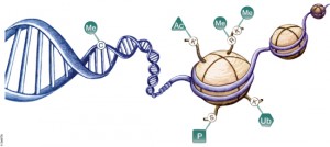
DNA is organized by protein:DNA complexes called nucleosomes in eukaryotes. Nucleosomes are composed of 147 base pairs of DNA wrapped around a histone octamer containing two copies of each core histone protein. Histone proteins play significant roles in many nuclear processes including transcription, DNA damage repair and heterochromatin formation. Histone proteins are extensively and dynamically post-translationally modified, and these post-translational modifications (PTMs) are thought to comprise a specific combinatorial PTM profile of a histone that dictates its specific function.  Abnormal regulations of PTM may lead to developmental disorders and disease development such as cancer.

Continue reading “Mass Spec Analysis of PTMs Using Minimal Sample Material”

Biotechnology From the Mouths of Babes

As a science writer, much of my day entails reviewing and revising marketing materials and technical literature about complex life science research products. I take for granted the understanding that I, my colleagues and our customers have of how these technologies work. This fact really struck me as I read an article about research to improve provider-patient communication in healthcare settings.

The researchers completed an analysis revealing that patient information materials had an average readability at a high school level, while the average patient reads at a fourth-grade level. These findings inspired the researchers to conduct a study in which they enlisted the help of elementary students to revise the content of the patient literature after giving them a short lesson on the material.

The resulting content did not provide more effective ways to communicate indications, pre- and post-op care, risks or procedures—that wasn’t really the point. Instead, the study underscores the important connection between patient literacy and health outcomes. More specifically, a lack of health literacy is correlated with poor outcomes and increased healthcare costs, prompting action from the US Department of Health & Human Services.

While healthcare information can be complex and full of specific medical terminology, I recognized that a lot of the technical and marketing information we create for our products at Promega has similar features. Wouldn’t it be interesting to find out how descriptions of some of our biggest technologies translate through the eyes and mouths of children?

After enlisting some help from my colleagues, I was able to catch a glimpse of how our complex technologies are understood by the little people in our lives. The parents and I explained a technology and then had our child provide a description or drawing of what they understood.

Continue reading “Biotechnology From the Mouths of Babes”

Optimized Detection of EPO-Fc in Human Biological Fluids

Recombinant erythropoietin (rhEPO) is often used as “doping agent” by athletes in endurance sports to increase blood oxygen capacity. Some strategies improve the pharmacological properties of erythropoietin (EPO) through the genetic and chemical modification of the native EPO protein. The EPO-Fcs are fusion proteins composed of monomeric or dimeric recombinant EPO and the dimeric Fc region of human IgG molecules. The Fc region includes the hinge region and the CH2 and CH3 domains. Recombinant human EPOs (rhEPO) fused to the IgG Fc domain demonstrate a prolonged half-life and enhanced erythropoietic activity in vivo compared with native or rhEPO.

Drug-testing agencies will need to obtain primary structure information and develop a reliable analytical method for the determination of EPO-Fc abuse in sport. The possibility of EPO-Fc detection using nanohigh-performance liquid chromatography−tandem mass spectrometry (HPLC−MS/MS) was already demonstrated (1). However, the prototyping peptides derived from EPO and IgG are not selective enough because both free proteins are naturally presented in human serum. In a recent publication, researchers describe the effort to identify peptides covering unknown fusion breakpoints (later referred to as “spacer” peptides; 2). The identification of “spacer” peptides will allow the confirmation of the presence of exogenous EPO-Fc in human biological fluids.

Are you looking for proteases to use in your research?
Explore our portfolio of proteases today.

A bottom-up approach and the intact molecular weight measurement of deglycosylated protein and its IdeS proteolytic fractions was used to determine the amino acid sequence of EPO-Fc. Using multiple proteases, peptides covering unknown fusion breakpoints (spacer peptides) were identified.

Results indicated that “spacer peptides” could be used in the determination of EPO-Fc fusion proteins in biological samples using common LC−tandem MS methods.

References

  1. Reichel, C. et al. (2012) Detection of EPO-Fc fusion protein in human blood: screening and confirmation protocols for sports drug testing.
    Drug Test. Anal. 4, 818−29.
  2. Mesonzhnik, N. et al. (2017) Characterization and Detection of Erythropoietin Fc Fusion Proteins Using Liquid Chromatography−Mass Spectrometry.
    J. of Proteome Res. 17, 689-97.

Shining Stars: Cool NanoLuc® Plasmid Constructs Available Through the Addgene Repository

Researchers having been sharing plasmids ever since there were plasmids to share. Back when I was in the lab, if you read a paper and saw an interesting construct you wished to use, you could either make it yourself or you could “clone by phone”.  One of my professors was excellent at phone cloning with labs around the world and had specific strategies and tactics for getting the plasmids he wanted. Addgene makes this so much easier to share your constructs from lab to lab. Promega supports the Addgene mission statement: Accelerate research and discovery by improving access to useful research materials and information.  Many of our technology platforms like HaloTag® Fusion Protein, codon-optimized Firefly luciferase genes (e.g., luc2), and NanoLuc® Luciferase are present in the repository. We encourage people to go to Addgene to get new innovative tools. Afterall, isn’t science better when we share?

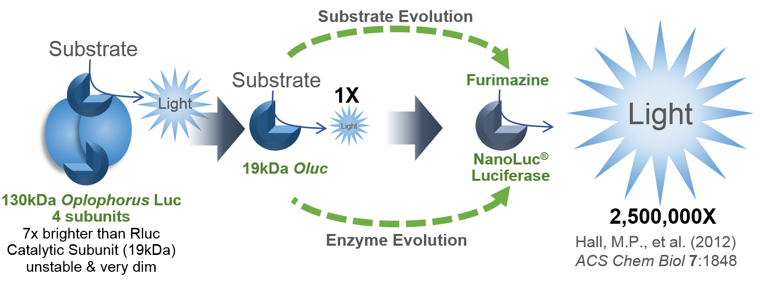
I’d like to focus on some tools in the Addgene collection based on NanoLuc® Luciferase (NLuc).  The creation of NanoLuc® Luciferase and the optimal substrate furimazine is a good story (1).  From a deep sea shrimp to a compact powerhouse of bioluminescence, NLuc is 100-fold brighter than our more common luciferases like firefly (FLuc) and Renilla (RLuc) luciferase.  This is important not so much for how bright you can make a reaction but for how sensitive you can make a reaction.  NLuc requires 100-fold less protein to produce the same amount of light from a Fluc or RLuc reaction.  NLuc lets you work at physiological concentrations.  NLuc is bright enough to detect endogenous tagged genes generated through the CRISPR/Cas9 knock-in.  NLuc is very inviting for endogenous tagging as it is only 19kDa.  An example is the CRISPaint-NLuc construct (Plasmid #67178) for use in the system outlined in Schmid-Burgk, J.L. et al (2).

Two applications of NanoLuc® Technology have caught my attention through coupling the luciferase with fluorescent proteins to make better imaging reporters and biosensors. Continue reading “Shining Stars: Cool NanoLuc® Plasmid Constructs Available Through the Addgene Repository”

Luciferase Immunoprecipitation System Assay (LIPS): Expression of Luciferase Antigen using TNT Transcription/Translation Kit

NanoLuc dual reporters
Illustration showing NanoLuc and firefly luciferase reporters.

The luciferase immunoprecipitation system (LIPS) assay is a liquid phase immunoassay allowing high-throughput serological screening of antigen-specific antibodies. The immunoassay involves quantitating serum antibodies by measuring luminescence emitted by the reporter enzyme Renilla luciferase (Rluc) fused to an antigen of interest. The Rluc-antigen fusion protein is recognized by antigen-specific antibodies, and antigen-antibody complexes are captured by protein A/G beads that recognize the Fc region of the IgG antibody (1).

In a recent publication (2), this assay was used to assess the presence of autoantibodies against ATP4A and ATP4B subunits of parietal cells H+, K+-ATPase in patients with atrophic body gastritis and in controls. Continue reading “Luciferase Immunoprecipitation System Assay (LIPS): Expression of Luciferase Antigen using TNT Transcription/Translation Kit”

Determination of Antibody Mechanism of Action Using IdeS Protease

Monoclonal antibodies (mAbs) have been widely used to eliminate undesired cells via various mechanisms, including antibody-dependent cell-mediated cytotoxicity (ADCC), complement-dependent cytotoxicity (CDC) and programmed cell death (PCD). Unlike the Fc-dependent mechanism of ADCC and CDC, certain antibody–antigen interactions can evoke direct PCD via apoptosis or oncosis. Previously, researchers have reported the specific killing of undifferentiated human embryonic stem cells (hESC) by mAb84 (IgM) via oncosis (1)

In a recent publication (2), a monoclonal antibody (mAb), TAG-A1 (A1), was generated to selectively kill residual undifferentiated human embryonic stem cells (hESC). One of the many experimental tools used to characterize the mechanism of oncosis was the fragmention of the A1 antibody with IdeS and papain.

Learn more about IdeS and IdeZ Protease available from Promega.

Papain digestion of IgG produces Fab fragments in the presence of reducing agent. F(ab)2 fragments of A1 were produced using IdeS Protease.

The results indicate that both Fab_A1 and F(ab)2_A1 bind to hESC but only F(ab)2_A1 retained hESC killing. Hence bivalency, but not Fc-domain, is essential for A1 killing on hESC.

  1. Choo, A.B. et al. (2008) Selection against undifferentiated human embryonic stem cells by a cytotoxic antibody recognizing podocalyxin-like protein-1. Stem Cells  26, 1454.
  2. Zheng, J.Y. et al. (2017) Excess reactive oxygen species production mediates monoclonal antibody-induced human embryonic stem cell death via oncosis. Cell Death and Differentiation 24, 546–58.

Are you looking for proteases to use in your research?
Explore our portfolio of proteases today.

Analysis of a biosimilar mAb using Mass Spectrometry

Several pharmaceutical companies have biosimilar versions of therapeutic mAbs in development. Biosimilars can promise significant cost savings for patients, but the unavoidable differences
between the original and thencopycat biologic raise questions regarding product interchangeability. Both innovator mAbs and biosimilars are heterogeneous populations of variants characterized by differences in glycosylation,oxidation, deamidation, glycation, and aggregation state. Their heterogeneity could potentially affect target protein binding through the F´ab domain, receptor binding through the Fc domain, and protein aggregation.

As more biosimilar mAbs gain regulatory approval, having clear framework for a rapid characterization of innovator and biosimilar products to identify clinically relevant differences is important. A recent reference (1) applied a comprehensive mass spectrometry (MS)-based strategy using bottom-up, middle-down, and intact strategies. These data were then integrated with ion mobility mass spectrometry (IM-MS) and collision-induced unfolding (CIU) analyses, as well as data from select biophysical techniques and receptor binding assays to comprehensively evaluate biosimilarity between Remicade and Remsima.

The authors observed that the levels of oxidation, deamidation, and mutation of individual amino acids were remarkably similar. they found different levels of C-terminal truncation, soluble protein aggregates, and glycation that all likely have a limited clinical impact.  Importantly, they identified more than 25 glycoforms for each product and observed glycoform population differences.

Overall the use of mass spectrometry-based analysis provides rapid and robust analytical information vital for biosimilar development. They demonstrated the utility of our multiple-attribute monitoring workflow using the model mAbs Remicade and Remsima and have provided a template for analysis of future mAb biosimilars.

1. Pisupati, K. et. al. (2017) A Multidimensional Analytical Comparison of Remicade and the Biosimilar Remsima. Anal. Chem 89, 38–46.