Characterization of Ubiquitination Using Cell-Free Expression

Ubiquitination refers to the post translational modification of a protein by attachment of one or more ubiquitin monomers. The most prominent function of ubiqutin is labeling proteins for proteasome degradation. In addition to this function ubiquitination also controls the stability, function and intracellular localization of a wide variety of proteins.
Cell free expression can be used to characterize ubiquitation of proteins. Target proteins are expressed in a rabbit reticulocyte cell free system (supplemented with E1 ubiquitin activating enzyme, E2 ubiquitin –conjugating enzyme, and ubiquitin). Proteins that have been modified can be analyzed by a shift in migration on polyacrylamide gels.

The following references illustrate the use of cell free expression for this application.

Jung, Y.S. et al. (2011) The p73 Tumor Suppressor Is Targeted by Pirh2 RING Finger E3 Ubiquitin Ligase for the Proteasome-dependent Degradation. J. Biol. Chem. 286, 35388–95.

Su, C-H, et al. (2010) 14-3-3sigma exerts tumor-suppressor activity mediated by regulation of COP1 stability. Cancer. Res. 71, 884–94.

Naoe, H. et al. (2010). The anaphase-promoting complex/cyclosome activator Cdh1 modulates Rho GTPase by targeting p190 RhoGAP for degradation. Mol. Cell. Biol. 30, 3994-05.

de Thonel, A. et al. (2010) HSP27 controls GATA-1 protein level during erythroid cell differentiation. Blood 116, 85–96.

Kaneko, M. et al. (2010) Loss of HRD1-mediated protein degradation causes amyloid precursor protein accumulation and amyloid-beta generation. J. Neurosci. 30, 3924–32.

Cell-Free Kinase Assays

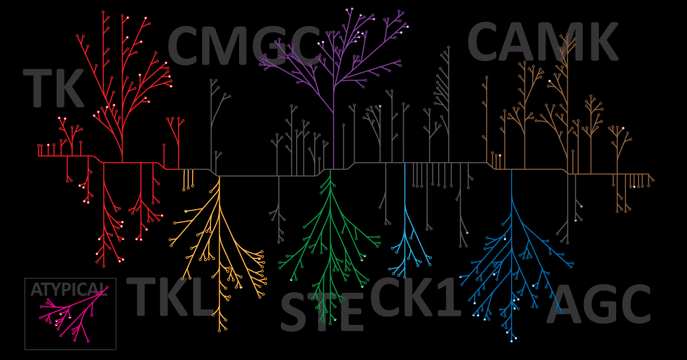
Protein phosphorylation is one of the most biologically relevant modifications and is involved in many eukaryotic and prokaryotic cellular signaling processes. It is estimated that one-third of human proteins are phosphorylated.

The following examples utilize the ability of cell free experession to express active proteins, and when supplemented with the necessary components (e.g., ATP, NaCl), to be used for the characterization of phosphorylation events.

Modrof, J. et al. (2005) Phosphorylation of bluetongue virus nonstructural protein 2 is essential for formation of viral inclusion bodies. J. Vir. 79, 10023–31. Use of TNT® cell-free to express NS2 and NS2 mutant proteins for use in vitro kinase assays to confirm phosphorylation by protein kinase CK2.

Kwon, S. et al. (2005) Signal pathway of hypoxia-inducible factor-1alpha phosphorylation and its interaction with von Hippel-Lindau tumor suppressor protein during ischemia in MiaPaCa-2 pancreatic cancer cells. Clin. Cancer Res. 11, 7607–13. The TNT® system was used to identify which p38 mitogen-activated protein kinase isoform(s) was cabable of phosphorylation of HIF—1 alpha

Harris, J. et al. (2006). Nuclear accumulation of cRel following C-terminal phosphorylation by TBK1/IKK epsilon. J. Immunol. 177, 2527–35. IKK and IKK mutants were expressed using TNT and used in a vitro kinase assay to characterize the recognition motif in cRel transcription domain

Jailais, Y. et al. (2011) Tyrosine phosphorylation controls brassinosteroid receptor activation by triggering membrane release of its kinase inhibitor. Genes Dev. 25, 232–37. Using a vitro kinase assay, full–length and truncations versions of the Brassinostediod-insentive receptor protein were expressed using the TNT® system and incubated with purified BR11 kinase domain to determine binding sites of the two proteins.

Optimized Protein Expression: Flexi Rabbit Reticulocyte Lysate

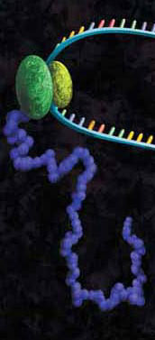
A protein chain being produced from a ribosome.

mRNAs commonly exhibit differing salt requirements for optimal translation. Small variations in salt concentration can lead to dramatic differences in translation efficiency. The Flexi® Rabbit Reticulocyte Lysate System allows translation reactions to be optimized for a wide range of parameters, including
Mg2+ and K+ concentrations and the choice of adding DTT. To help optimize Mg2+ for a specific message, the endogenous Mg2+ concentration of each lysate batch is stated in the product information included with this product.

The following references utilize the features of Flexi Rabbit Reticulocyte Lysate System to investigate certain parameters of translation:

Vallejos, M. et al. (2010)The 5′-untranslated region of the mouse mammary tumor virus mRNA exhibits cap-independent translation initiation. Nucl Acids Res. 38, 618–32. Identification of internal ribosomal ribosomal entry site in the 5’ untranslated region of the mouse mammary tumor virus mRNA.

Spriggs, K. et al. (2009) The human insulin receptor mRNA contains a functional internal ribosome entry segment. Nucl. Acids. Res. 17, 5881–93. Identification of a functional internal ribosome entry site in the human insulin receptor mRNA.

Powell, M. et al. (2008) Characterization of the termination-reinitiation strategy employed in the expression of influenza B virus BM2 protein. RNA 14, 2394–06. Analysis of the mRNA signals involved in the expression of influenza B virus BM2 protein.

Sato, V. et al. (2007) Measles virus N protein inhibits host translation by binding to eIF3-p40. J. Vir. 81, 11569–76. Charaterized the effect of the measles virus N protein binding to the translation initiation factor eIF3-p40 on the expression of various proteins in rabbit reticulocyte lysate.

Hirao, K. et al. (2006) EDEM3, a soluble EDEM homolog, enhances glycoprotein endoplasmic reticulum-associated degradation and mannose trimming. J. Biol. Chem. 281, 9650–58. The EDEM3 protein was expressed in the presence of canine microsomal membranes to establish that co-translational translocation occurs into the endoplasmic reticulum.

Shenvi, C. et al. (2005) Accessibility of 18S rRNA in human 40S subunits and 80S ribosomes at physiological magnesium ion concentrations–implications for the study of ribosome dynamics. RNA 11, 1898–08. Characterization of ribosome dynamics under different ionic conditions.

Nair, A. et al. (2005) Regulation of luteinizing hormone receptor expression: evidence of translational suppression in vitro by a hormonally regulated mRNA-binding protein and its endogenous association with luteinizing hormone receptor mRNA in the ovary. J. Biol. Chem. 280, 42809–16. Examined the affect of luteinizing hormone receptor mRNA binding protein on transltional suppression of luteinizing hormone receptor RNA.

Optimization of Western Blots Detecting Proteins Synthesized Using Cell-Free Expression #2

Detection of protein expressed using cell-free systems is required for most applications such as protein:protein interaction and protein:nucleic acid interaction studies. Traditionally, one adds radioactive [35S]methionine to cell-free expression reactions, and the methionine is incorporated into the expressed protein, allowing detection by autoradiography. Many researchers are moving away from radioactivity. Traditional Western blot analysis provides the researcher a nonradioactive method for detection but, if performed improperly, can result in high background, which can mask expressed proteins and affect downstream applications.

One critical step in producing low-background, high-signal Western blots is choosing the correct dilution of the primary antibody. Typically the manufacturer recommends antibody dilution from 1:1,000 to 1:2,500 for standard western blotting experiments. However when using crude lysates as a source of the target protein, these recommendations exhibit significant background. When the antibody was diluted 1:50,000, background was decreased significantly, and the positive signal was a large percentage of the total signal.

As a general recommendation when performing Western blot analysis of proteins expressed in cell-free systems, one must experimentally determine the optimal dilution of the primary antibody. In the Western blots performed in this study, primary antibodies were diluted ~50-fold more than the provider’s recommended dilution.

For additional technical details refer to this recent article published in Promega’s PubHub:

Hook, B and Schagat, T. (2011) Non-Radioactive Detection of Proteins Expressed in Cell-Free Expression Systems Promega Corporation Web site. Accessed August 17, 2011.

Cell-Free Applications: RNA Toeprinting

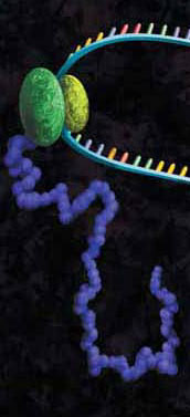
A protein chain being produced from a ribosome.
Precise mapping of the positions of ribosomes and associated factors on mRNAs is essential for characterizing the mechanism of translation. Using the toeprinting assay, mRNA is translated using purified components or crude cell lysates such as rabbit reticulocyte. Cycloheximide is added to the reaction to inhibit elongation. This arrests the position of the ribosomes on the mRNA transcript. The mRNA/ribosomal complex are then copied into cDNA by reverse transcriptase using a complementary radiolabeled primer. Where the reverse transcriptase meets the ribosome bound to the mRNA, cDNA extension is halted, and a toeprint cDNA fragment is generated.

The following references use rabbit reticulocyte lysates as the basis for toeprinting experiments to better understand the mechanism of translation.

Weill, L. et al. (2010)Nucl. Acid, Res. 38, 1367–81. A combination of chemical/enzymatic analyses indicated that gag open reading frame of three viruses adopts a stable secondary structure that allows IRES mediated translation. Mutations that destabilized conserved elements severely inhibit translation. Additional analysis via toeprinting showed HIV-2 IRES has the unique ability to attract up to three initiation complexes on a single RNA molecule.

De Breyne, S. et al. (2008) RNA 14, 367–80. The Simian picornavirus type 9 (SPV9) genome contains a group of IRES that resembles hepacivirus/pestvirus (HP) IRES. Characterization of the initiation process using the toeprinting assay in correlation with other techniques revealed aspects that resemble initiation on the HP IRES and others that are unique to SPV9.

Andreev, D. et al. (2008) RNA 14, 233–39. Rel E is a well characterized toxin involved in the nutritional stress response in bacteria and archae. Rel lacks any eukaryote homolog. Based on toeprinting data, it was demonstrated that RelE cleaves mRNA in the A site of the eukaryote ribosome.

Cell-Free Expression: Non-Radioactive Detection/Applications

The Transcend™ Non-Radioactive Translation Detection Systems allow nonradioactive detection of proteins synthesized using cell free expression. Using these systems, biotinylated lysine residues are incorporated into nascent proteins during translation, This biotinylated lysine is added to the translation reaction as a precharged ε-labeled biotinylated lysine-tRNA complex rather than a free amino acid. After SDS-PAGE and electroblotting, the biotinylated proteins can be visualized by binding either Streptavidin-Alkaline Phosphatase (Streptavidin-AP) or Streptavidin-Horseradish Peroxidase (Streptavidin-HRP), followed either by colorimetric or chemiluminescent detection. Typically, these methods can detect 0.5–5ng of protein within 3–4 hours after gel electrophoresis and can be used for a variety of proteomics related applications. Examples include: Continue reading “Cell-Free Expression: Non-Radioactive Detection/Applications”

Cell-Free Applications:Protein Arrays (Nucleic Acid Programmable)

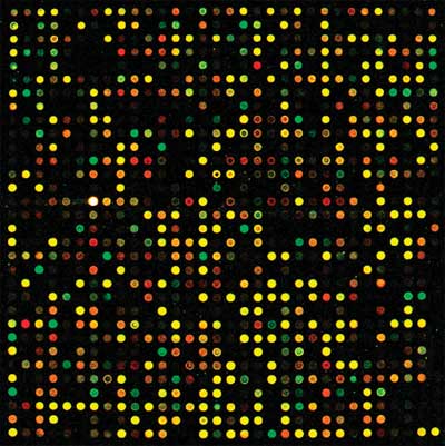
The traditional methods of generating protein arrays require the separate expression of hundreds of proteins, followed by purification and immobilization of the proteins on a solid surface. Cell-Free protein array technology produces protein microarrays by performing in vitro synthesis of the target protein from their DNA templates.
One approach for the generation of cell- free based microarrays is the nucleic acid programmable protein array (NAPPA).

NAPPA uses DNA template that is biotinylated and is bound to avidin that is pre-coated onto the protein capture surface. Newly synthesized proteins which are tagged with GST are then immobilized next to the template DNA by binding to an adjacent polyclonal anti-GST capture antibody. The following references illustrate the use of NAPPA to screen hundreds of proteins. Continue reading “Cell-Free Applications:Protein Arrays (Nucleic Acid Programmable)”

Cell-Free Protein Expression: Characterization of Plant Proteins

Cell free protein expression can be utilized for the analysis of: protein/protein interactions, protein nucleic acid interactions, analysis of post translational modifications and many other applications. The majority of these references are based on the characterization of mammalian proteins.
However there are several references using TNT-based systems (either rabbit reticulocyte lysate or wheat germ based) for the analysis of proteins from plants, examples include: Continue reading “Cell-Free Protein Expression: Characterization of Plant Proteins”

Use of Cell-Free Protein Expression for Epigenetics-Related Applications

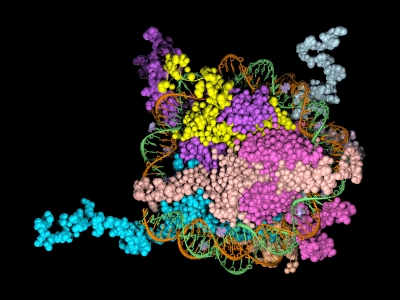
Epigenetics is the study of the processes involved in the genetic development of an organism, especially the activation and deactivation of genes. One way that genes are regulated is through the remodeling of chromatin. Chromatin is the complex of DNA and the histone proteins with which it associates. The conformation of chromatin is profoundly influenced by the post-translational modification of the histone proteins. These modifications include acetylation, methylation, ubiquitylation, phosphorylation and sumolyation. The following references illustrate the use of cell-free expression to characterize this process.

Shao, Y. et al. (2010) Nucl. Acid. Res. 38, 2813–24.
Carbonic anhydrase IX (CAIX) plays an important role in the growth and survival of tumor cells.The MORC proteins contain a CW-type zinc finger domain and are predicted to have the function of regulating transcription, but no MORC2 target genes have been identified. CAIX mRNA to be down-regulated 8-fold when MORC2 was overexpressed. Moreover, MORC2 decreased the acetylation level of histone H3 at the CAIX promoter. Among the six HDACs tested, histone deacetylase 4 (HDAC4) had a much more prominent effect on CAIX repression. Assays showed that MORC2 and HDAC4 were assembled on the same region of the CAIX promoter. Interaction between MORC2 and HDAC 4 were confirmed by using cell free expression of MORC2 and GST-HDAC (GST pull-downs). Cell-free expression was also used to express MORC2 proteins to determine through gel shifts the binding location on the CAIX promoter region (gel shift experiments)

Denis, H. et al. (2009) Mol. Cell. Biol. 29, 4982–93.
The recent identification of enzymes that antagonize or remove histone methylation offers new opportunities to appreciate histone methylation plasticity in the regulation of epigenetic pathways. PAD4 was the first enzyme shown to antagonize histone methylation. Very little is known as to how PADI4 silences gene expression. Through the use of cell-free expression to express both PAD4 and HDAC1 proteins and E. coli expression of GST fusions of PAD4 and HDAC1, pulldown experiments confirmed by in vivo experiments that PADI4 associates with the histone deacetylase 1 (HDAC1), and the corresponding activities, associate cyclically and coordinately with the pS2 promoter during repression phases.

Brackertz, M. et al. (2006) Nucl. Acid. Res. 34, 397-406.
The Mi-2/NuRD complex is a multi-subunit protein complex with enzymatic activities involving chromatin remodeling and histone deacetylation. The function of p66α and of p66β within the multiple subunits has not been addressed. GST-fused histone tails of H2A, H2B, H3 and H4 were expressed in E. coli used in an in vitro pull-down assay with radioactively labeled p66-constructs expressed using cell free systems. Deletions at the C terminus noted reduced binding of p66 where as deletions at the N terminus did not affect binding. Also observed was that acetylation of histone tails reduces the association with both p66-proteins in vitro.

Zhou, R. et al. (2009) Nucl. Acids. Res. 37, 5183–96.
Lymphoid specific helicase (Lsh) belongs to the family of SNF2/helicases. Disruption of Lsh leads to developmental growth retardation and premature aging in mice. However, the specific effect of Lsh on human cellular senescence remains unknown. In vivo results noted that Lsh requires histone deacetylase (HDAC) activity to repress p16INK4a. Moreover, overexpression of Lsh is correlated with deacetylation of histone H3 at the p16 promoter. In vitro pull-downs using cell free expression and GST fusions from E. coli were used to collaborate interactions between Lsh, histone deacetylase 1 (HDAC1) and HDAC2 observed in vivo.

Purification of biotinylated proteins

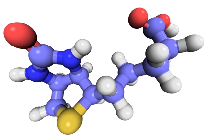
Biotinylation is an attractive approach for protein complex purification due to the very high affinity (Kd = 10–15 M) of avidin/streptavidin for biotinylated templates. With typical avidin or streptavidin, the biotin-binding affinity is so great that purification with these traditional media require denaturing conditions for elution,such as 8 M Guanidine•HCl at pH 1.5 or boiling in reducing SDS-PAGE sample loading buffer. To avoid these harsh conditions SoftLink™ Soft Release Avidin resin can be used. These particles consist of monomeric avidin coupled to a methylacrylate resin.

This resin provides the same specificity of binding to biotin afforded by tetrameric biotin, but enables the release of biotinlylated molecules under mild nondenaturing conditions (5mM biotin).

The following are recent references that used the SoftLink™ Resin for the noted application:

Kashwayama, Y. et al. (2010) Identification of a substrate-binding site in a peroxisomal beta-oxidation enzyme by photoaffinity labeling with a novel palmitoyl derivative. J. Biol. Chem. 285, 26315–25. (Purification of photoaffinity labeled proteins for subquenant binding/activity experiments)

Takahashi, M. et al. (2010) Tailor-made RNAi knockdown against triplet repeat disease-causing alleles. Proc. Natl. Acad. Sci. 107, 21731-36 (Innovated procedure using biotin labeled cDNAs for the identification of nucleotide variations)

Kress, D. et al. (2009) An asymmetric model for Na+-translocating glutaconyl-CoA decarboxylases. J. Biol.Chem. 284, 28401–9 (Purification of Clostridium biotin carrier proteins that play a role in decarboxylation)

Akahori, Y. et al. (2009) Characterization of neutralizing epitopes of varicella-zoster virus glycoprotein H. J. Virol. 83, 2020–4. (purification of double stranded cDNA fragments amplified by PCR with a biotin-tagged PCR primer)

Shonsey, E.M. et al. (2008) Inactivation of human liver bile acid CoA:amino acid N-acyltransferase by the electrophilic lipid, 4-hydroxynonenal. J. Lipid Res. 49, 282–94 (purification of recombinant protein expressed in E.coli containing C-terminal avidin tag)

Andachi, Y. et al. (2008) A novel biochemical method to identify target genes of individual microRNAs: identification of a new Caenorhabditis elegans let-7 target. RNA 14, 2440–51 (purification of double stranded cDNA fragments amplified by PCR with a biotin-tagged PCR primer)

For more information about SoftLink™ Soft Release Avidin Resin, please visit our website.