Another Step Closer to Understanding Epigenetic Gene Regulation

Chromatin fiber

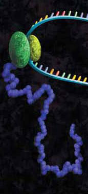
Back when I was a graduate student (more than a few years ago), I remember hearing another student joke that if a member of his thesis committee asked him to explain an unexpected or unusual result, he was going to “blame” epigenetics. At that time, the study of epigenetic gene regulation was in its infancy, and scientists had much to learn about this mysterious regulatory process. Fast forward to today, and you’ll find that scientists know a lot more about basic epigenetic mechanisms, although there is still plenty to learn as scientists discover that the topic is much more complicated than initially thought, as is often the case in science. A recent EMBO Journal article is contributing to our knowledge by shedding light on the role of the TET family of DNA-modifying enzymes in epigenetics (1).

Continue reading “Another Step Closer to Understanding Epigenetic Gene Regulation”

Proteinase K: An Enzyme for Everyone

protein expression purification and analysis

We recently posted a blog about Proteinase K, a serine protease that exhibits broad cleavage activity produced by the fungus Tritirachium album Limber. It cleaves peptide bonds adjacent to the carboxylic group of aliphatic and aromatic amino acids and is useful for general digestion of protein in biological samples. In that previous blog we focused on its use to remove RNase and DNase activities. However, the stability of Proteinase K in urea and SDS and its ability to digest native proteins make it useful for a variety of applications. Here we provide a brief list of peer-reviewed citations that demonstrate the use of proteinase K in DNA and RNA purification, protein digestion in FFPE tissue samples, chromatin precipitation assays, and proteinase K protection assays:

Continue reading “Proteinase K: An Enzyme for Everyone”

PNGase F, a Novel Endoglycosidase

11123MA

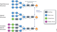
PNGase F (Cat.# V4831) is a recombinant glycosidase cloned from Elizabethkingia meningoseptica and overexpressed in E. coli, with a molecular weight of 36kD.

PNGase F catalyzes the cleavage of N-linked oligosaccharides between the innermost GlcNAc and asparagine residues of high mannose, hybrid, and
complex oligosaccharides from N-linked glycoproteins. PNGase F will not remove oligosaccharides containing alpha-(1,3)-linked core fucose,
commonly found on plant glycoproteins.

Applications
Determining whether a protein is in fact glycosylated is the initial step in glycoprotein analysis. Polyacrylamide gel electrophoresis in the
presence of sodium dodecyl sulfate (SDS-PAGE) has become the method of choice as the final step prior to mass spec analysis. Glycosylated proteins often migrate as diffused bands by SDS-PAGE. A marked decrease in band width and change in migration position after treatment with PNGase F is considered evidence of N-linked glycosylation.

Gel based data are often correlated with information obtained from mass spec analysis. Asn-linked type glycans can be cleaved enzymatically by PNGase F yielding intact oligosaccharides and a slightly modified protein in which Asn residues at the site of de-N-glycosylation are converted to Asp, by converting the previously carbohydrate-linked asparagine into an aspartic acid, a monoisotopic mass shift of 0.9840Da is observed. The deglycosylated peptides are then analyzed by tandem mass spectrometry (MS/MS), and software algorithms are used to correlate the experimental fragmentation spectra with theoretical tandem mass spectra generated from peptides in a protein database.

Enhancing Proteomics Data Using Arg-C Protease

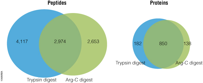
Arg-C (clostripain), Sequencing Grade (Cat.# V1881), is a specific endoproteinase isolated from the soil bacterium Clostridium histolyticum. It preferentially cleaves at the C-terminal side of arginine (R) residues. Unlike trypsin, Arg-C efficiently cleaves arginine sites followed by proline (P). This difference is important because every twentieth arginine is followed by proline. To illustrate this benefit, Arg-C was evaluated for protein analysis in two different experiments. In the first experiment, we studied the use of Arg-C for proteomic analysis. Yeast provides an excellent model proteome because its genome is well annotated. Yeast extract was digested in two parallel reactions, using trypsin in the first reaction and Arg-C in the second, using a conventional protocol consistent with LC-MS/MS analysis. As expected the trypsin digestion resulted in a high number of peptide and protein identifications (Figure 1). However, many peptides remained elusive. The parallel Arg-C digestion complemented the trypsin digestion by recovering an additional 2,653 peptides and providing a 37.4% increase in the number of identified peptides. Digesting with Arg-C also resulted in an increase in the number of identified proteins. In fact, 138 new proteins were identified in Arg-C digest compared to the parallel trypsin digest, offering a 13.4% increase in the overall number of identified proteins.

Figure 1. Side-by-side analysis of trypsin-digested and Arg-C digested yeast proteins.

Are you looking for proteases to use in your research?
Explore our portfolio of proteases today.

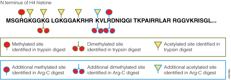
In a second experiment, the ability of Arg-C to analyze individual proteins was analyzed, selecting human histone H4 as a model protein. Like other histones, this protein is heavily modified post translational modifications (PTMs) that alter histone structure and regulate interaction with transcription factors. As a result, histone PTMs are implicated in gene regulation and associated with multiple disorders. Technical challenges, however, impede histone PTM analysis. Histone PTMs are complex and some, such as acetylation and methylation, prevent trypsin digestion, as shown by our data. In this experiment, trypsin digestion of histone H4 identified several PTMs (Figure 2). However, certain PTMs were missing. By digesting histone H4 with Arg-C, we were able to identify the missing PTMs including mono-, dimethylated and acetylated lysine and arginine residues. We speculate that the PTMs in human histone H4, which modified arginine and lysine residues, rendered trypsin unsuitable for preparing the corresponding histone regions for mass spectrometry. The problem was rectified by replacing trypsin with Arg-C.

Figure 2. Identification of histone h4 PTMs after Arg-C digestion.

Endo H Application: Monitoring Protein Trafficking

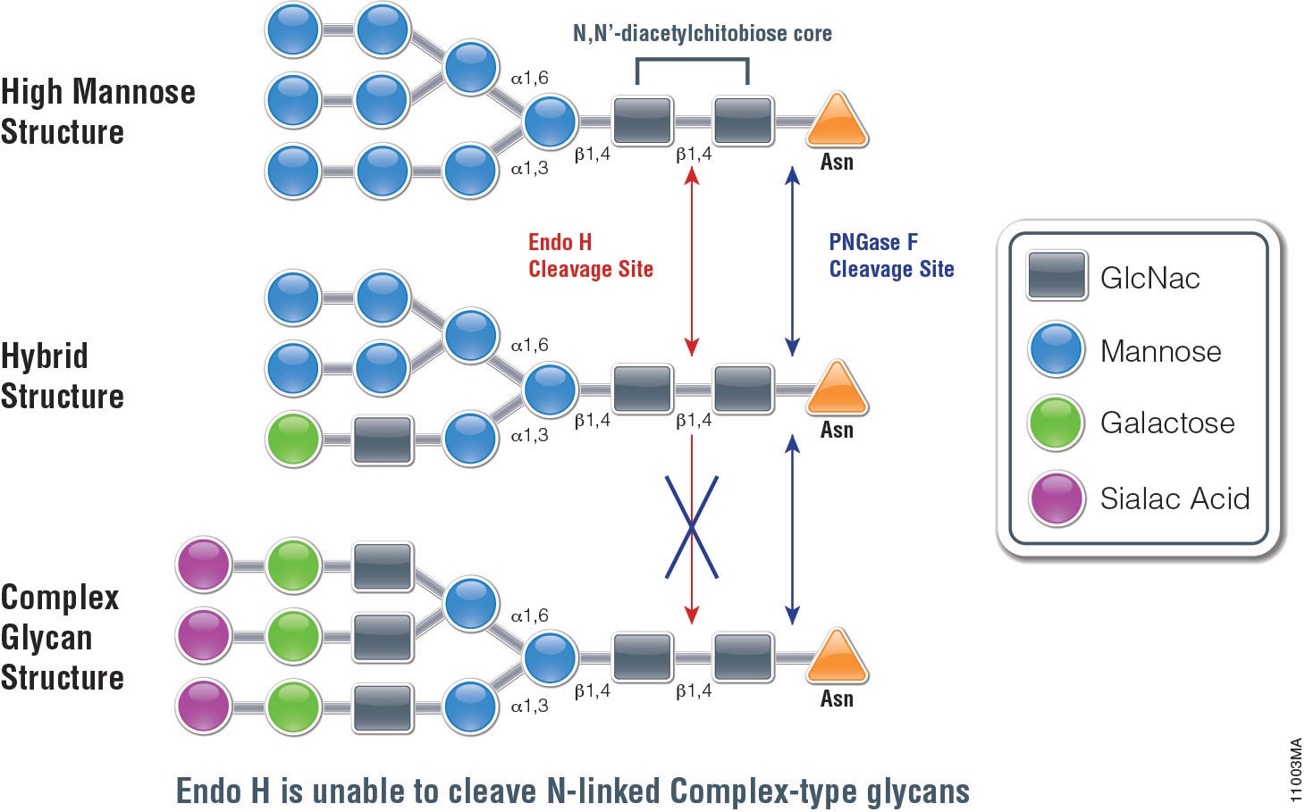
Endo H (Endo-Ăź-N-acetylglucosaminidase H) is a 29,000 dalton protein with optimal activity between pH 5 and 6. In contrast to PNGase F, which cleaves all N-linked glycans at the site of attachment to Asparagine (Asn), (with the exception of those with fucose attached to the core GlcNac moieties), Endo H hydrolyses the bond connecting the two GlcNac groups that comprise the chitobiose core (see Figure 1.). In addition, Endo H cleaves high mannose and hybrid glycans, whereas complex glycans (those with more than 4 different sugar types per glycan chain, including the GlcNac groups) are resistant to hydrolysis.

The unique specificty of Endo H and PNGase F can be used to monitor protein trafficking. Basic N-Glycosylation occurs in the endoplasmic reticulum. Proteins in this stage are sensitive to Endo H digestion. If proteins have entered the Golgi body where additional modifications occur to the glycan, they are resistant to Endo H digestion.

The following references illustrate this application:

Cell free application: Sumoylation characterization

Small Ubiquitin-like Modifier (or SUMO) proteins are a family of small proteins that are covalently attached to and detached from other proteins in cells to modify their function. SUMOylation is a post-translational modification involved in various cellular processes, such as nuclear-cytosolic transport,
transcriptional regulation, apoptosis, protein stability and response to stress.

In contrast to ubiquitin, SUMO is not used to tag proteins for degradation. Mature SUMO is produced when the last four amino acids of the C-terminus have been cleaved off to allow formation of an isopeptide bond between the C-terminal glycine residue of SUMO and an acceptor lysine on the target protein.

Cell free expression can be used to characterize sumoylation of proteins. Target proteins are expressed in a rabbit reticulocyte cell free system (supplemented with necessary addition components,). Proteins that have been modified can be analyzed by a shift in migration on polyacrylamide gels, when compared to control reactions.

The following references illustrate the use of cell free expression for this application.

Brandl, A. et al. (2012) Dynamically regulated sumoylation of HDAC2 controls p53 deacetylation and restricts apoptosis following genotoxic stress. J. Mol. Cell. Biol. (online only)

Janer, A. et al. (2010). SUMOylation attenuates the aggregation propensity and cellular toxicity of the polyglutamine expanded ataxin-7. Human. Mol. Gen. 19, 181—95.

Rytinki, M. et al. (2009) SUMOylation attenuates the function of PGC-1alpha. J. Biol. Chem. 284, 26184-93.

Klein, U. et al. (2009) RanBP2 and SENP3 function in a mitotic SUMO2/3 conjugation-deconjugation cycle on Borealin. Mol. Cell. Biol. 20, 410–18.

Seo, W. and Ziltener, H. (2009) CD43 processing and nuclear translocation of CD43 cytoplasmic tail are required for cell homeostasis. Blood, 114, 3567–77.

Using Pepsin to Prepare F(ab’)2 Fragments and Determine Deuterium Exchange

Using Pepsin to Prepare F(ab’)2 Fragments

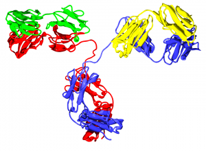
Pepsin is commonly used in the preparation of F(ab’)2 fragments from antibodies. In some assays, it is preferable to use only the antigen-binding (Fab) portion of the antibody. For these applications, antibodies may be enzymatically digested to produce an F(ab’)2 fragment of the antibody. To produce an F(ab’)2 fragment, IgG is digested with pepsin, which cleaves the heavy chains near the hinge region. One or more of the disulfide bonds that join the heavy chains in the hinge region are preserved, so the two Fab regions of the antibody remain joined together, yielding a divalent molecule (containing two antibody binding sites), hence the designation F(ab’)2. The light chains remain intact and attached to the heavy chain. The Fc fragment is digested into small peptides.

Continue reading “Using Pepsin to Prepare F(ab’)2 Fragments and Determine Deuterium Exchange”

High-Yield Cell-Free Protein Expression: Prokaryotic Based

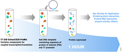
S30 E coli high yield extract schematicMany applications require amounts of protein that cannot be obtained using a eukaryotic cell-free expression system. As an alternative, a prokaryotic system can be used when this need arises. The E. coli S30 T7 High-Yield Protein Expression System is designed to express up to 500ÎĽg/ml of protein in 1 hour from plasmid vectors containing a T7 promoter and a ribosome binding site. The protein expression system provides an extract that contains T7 RNA polymerase for transcription and is deficient in OmpT endoproteinase and lon protease activity. All other necessary components in the system are optimized for protein expression. This results in greater stability and enhanced expression of target proteins.The following references highlight the use of this system for a variety of unique applications:

Loh, E. et al. (2011) An unstructured 5′-coding region of the prfA mRNA is required for efficient translation. Nuc. Acids. Res. (online) Examines the effect of upstream codon sequence/length on the correct ribosome binding and translation initiation of the pfrA protein.

Mitsuhashi, H. et al. (2010) Specific phosphorylation of Ser458 of A-type lamins in LMNA-associated myopathy patients. J. Cell. Sci. 123, 3893–900 By creating a series of mutations in the protein lamin A, Akt1 phosphorylation sites were determined.

Halvorsen, E. et al. (2011) Txe, an endoribonuclease of the enterococcal Axe-Txe toxin-antitoxin system, cleaves mRNA and inhibits protein synthesis. Microbiology 157, 387–97. S30 High Yield System was used to characterize the inhibitory effect of Txe toxin on protein expression.

Mo, P. et al. (2010) MDM2 mediates ubiquitination and degradation of activating transcription factor 3. J. Biol. Chem. 285, 26908–15. By using in vitro pull down experiments the researchers characterized the binding of AFT3 to MDM2 leading to the proteolysis of AFT3 system by ubiquitination.

Use of Nonspecific Proteases for Analysis of Proteins by Mass Spectrometry

mass spectrometry results

One of the approaches to identify proteins by mass spectrometry includes the separation of proteins by gel electrophoresis or liquid chromatography. Subsequently the proteins are cleaved with sequence-specific endoproteases. Following digestion the generated peptides are investigated by determination of molecular masses or specific sequence. For protein identification the experimentally obtained masses/sequences are compared with theoretical masses/sequences compiled in various databases.

Are you looking for proteases to use in your research?
Explore our portfolio of proteases today.

Nonspecific proteases such as pepsin, proteinase K, elastase and thermolysin can offer an alternative to traditional sequence-specific proteases for certain applications. The following references illustrate the use of nonspecific proteinases for the mass spec analysis of proteins:

Papasotiriou, D. et al. (2010) Peptide mass fingerprinting after less specific in-gel proteolysis using MALDI-LTQ-Orbitrap and 4-chloro-alpha-cyanocinnamic acid. J. Proteome. Res. 9, 2619–29. This reference demonstrates the use of either chymotrypsin, elastase, trypsin or proteinase K in combination with matrix CHCA for increase peptide identification and sequence coverage using MALDI.

Neue, K. et al. (2011) Elucidation of glycoprotein structures by unspecific proteolysis and direct nanoESI mass spectrometric analysis of ZIC-HILIC-enriched glycopeptides. J. Proteome. Res. 10, 2248–60. Notes use of thermolysin or elastase in combination with ZIC-HILIC enrichment as alternative method for the characterization of glycopeptides.

Baeumlisberger, D et al. (2011) Simple dual-spotting procedure enhances nLC-MALDI MS/MS analysis of digests with less specific enzymes. J. Proteome. Res. 10, 2889–94. Data noted that samples digested with elastase followed by nLC separation and subsequent alternative spotting on both MALDI-LTQ-Orbitrap and MALDL-TOF/TOF instruments resulted in 32% additional peptides.

Characterization of Ubiquitination Using Cell-Free Expression

Ubiquitination refers to the post translational modification of a protein by attachment of one or more ubiquitin monomers. The most prominent function of ubiqutin is labeling proteins for proteasome degradation. In addition to this function ubiquitination also controls the stability, function and intracellular localization of a wide variety of proteins.

Cell free expression can be used to characterize ubiquitation of proteins. Target proteins are expressed in a rabbit reticulocyte cell free system (supplemented with E1 ubiquitin activating enzyme, E2 ubiquitin –conjugating enzyme, and ubiquitin). Proteins that have been modified can be analyzed by a shift in migration on polyacrylamide gels.

The following references illustrate the use of cell free expression for this application.

Jung, Y.S. et al. (2011) The p73 Tumor Suppressor Is Targeted by Pirh2 RING Finger E3 Ubiquitin Ligase for the Proteasome-dependent Degradation. J. Biol. Chem. 286, 35388–95.

Su, C-H, et al. (2010) 14-3-3sigma exerts tumor-suppressor activity mediated by regulation of COP1 stability. Cancer. Res. 71, 884–94.

Naoe, H. et al. (2010). The anaphase-promoting complex/cyclosome activator Cdh1 modulates Rho GTPase by targeting p190 RhoGAP for degradation. Mol. Cell. Biol. 30, 3994-05.

de Thonel, A. et al. (2010) HSP27 controls GATA-1 protein level during erythroid cell differentiation. Blood 116, 85–96.

Kaneko, M. et al. (2010) Loss of HRD1-mediated protein degradation causes amyloid precursor protein accumulation and amyloid-beta generation. J. Neurosci. 30, 3924–32.