Cell-free expression application: Screening for successful oligo-mediated knockdown design

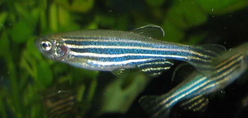
800px-ZebrafischAlthough previous references have provided data regarding the potential oncogenic role of the gene ETV7, there has been minimal investigation as to its physiological role.
In the following reference, Quintana, A. et al. (2014) Disease Models & Mechanisms 7, 265–70, zebrafish were used as in vivo model system to characterize ETV7.

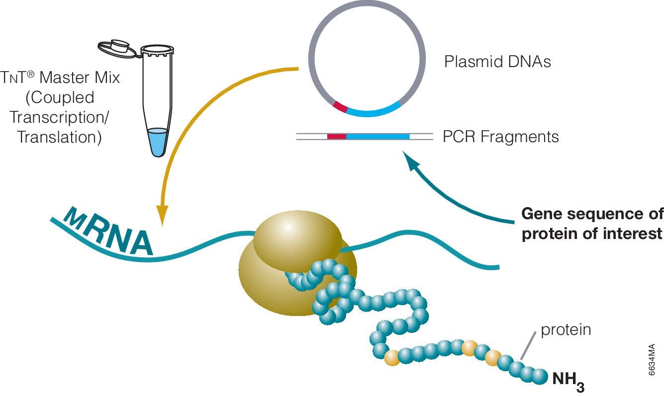
One key experiment required the morpholino-oligonucleotide -mediated knockdown of in vivo ETV7. Two independent morpholinos were designed: one that inhibited translation and the other that inhibited proper splicing of exon 3. The efficacy of the translation –blocking morpholino was assessed with cell free expression of ETV7-tagged with hemagglutinin (HA).

Western blot performed with anti-HA antibodies determined the extent of the knockdown compared to a control containing no morpholino added. Once an efficient design was determined via cell-free expression screening, it was used for in vivo experiments. In conjunction additional other techniques, concluded that ETV7 is essential for normal red blood cell development.

Cell-free Expression: A System for Every Need

6634MA

Cell-free protein expression is a simplified and accelerated avenue for the transcription and/or translation of a specific protein in a quasi cell environment. An alternative to slower, more cumbersome cell-based methods, cell-free protein expression methods are simple and fast and can overcome toxicity and solubility issues sometimes experienced in traditional E. coli expression systems. Continue reading “Cell-free Expression: A System for Every Need”

Novel Application for ProteaseMAX Surfactant: Cell Lysis

ProteaseMax Surfactant


The novel mass spectrometry compatible surfactant sulfonate-(sodium 3-((1-(furan-2-yl)undecyloxy) carbonylamino)-propane-1-sulfonate (i.e.ProteaseMAX) facilitates both in-gel and in-solution digestion applications by reducing the time required, enabling protein solubilization/denaturation and increasing peptide/protein identifications.

A new application was highlighted in a recent publication (1) which utilized ProteaseMAX to lyse cells prior to trypsin digestion and subsequent mass spec analysis. The composition of the buffer determines the overall efficiency of cell lysis, dissociation of protein complexes, protein solubility and ease of removal prior to LC/MS-MS analysis.

Are you looking for proteases to use in your research?
Explore our portfolio of proteases today.
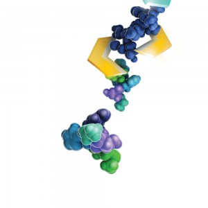
When compared to lysis buffers containing either urea or SDC, ProteaseMAX provided the optimal number of identified peptides/proteins.
In addition it can be easily removed from the lysate by acidic precipitation.

Reference

  1. Pirmoradian, M. et al. (2013). Rapid and deep human proteome analysis by single-dimension shotgun proteomics. Mol. Cell. Prot. 12, 3330–8.

ProTEV Protease Compound Compatibility Analysis

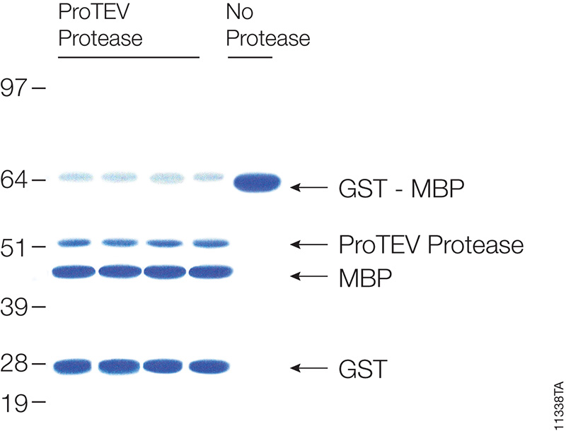
Cleavage of 20µg of GST-MBP fusion protein with ProTEV protease after 60 minutes at 30°C.

Many proteins are expressed as fusion partners with affinity tags, such as the HaloTag® fusion, glutathione-S-transferase (GST) or maltose binding protein (MBP), to selectively bind the proteins using affinity purification resins. While such resins yield high-purity protein quickly, the large affinity tags are undesirable for some downstream applications. Most expression vectors are designed with a specific protein cleavage site between the two fusion partners to remove the affinity tag after purification. ProTEV Protease recognizes a rare amino acid sequence, EXXYXQ, where X is any amino acid, and cleaves after the glutamine residue.

ProTEV Plus functions over a broad pH and temperature range. In a recent study the enzymatic activity of ProTEV Plus in the presence of various compounds (Table 1) commonly found in protein purification protocols were evaluated.

Continue reading “ProTEV Protease Compound Compatibility Analysis”

Trypsin/Lys-C Mix: Alternative for standard trypsin protein digestions

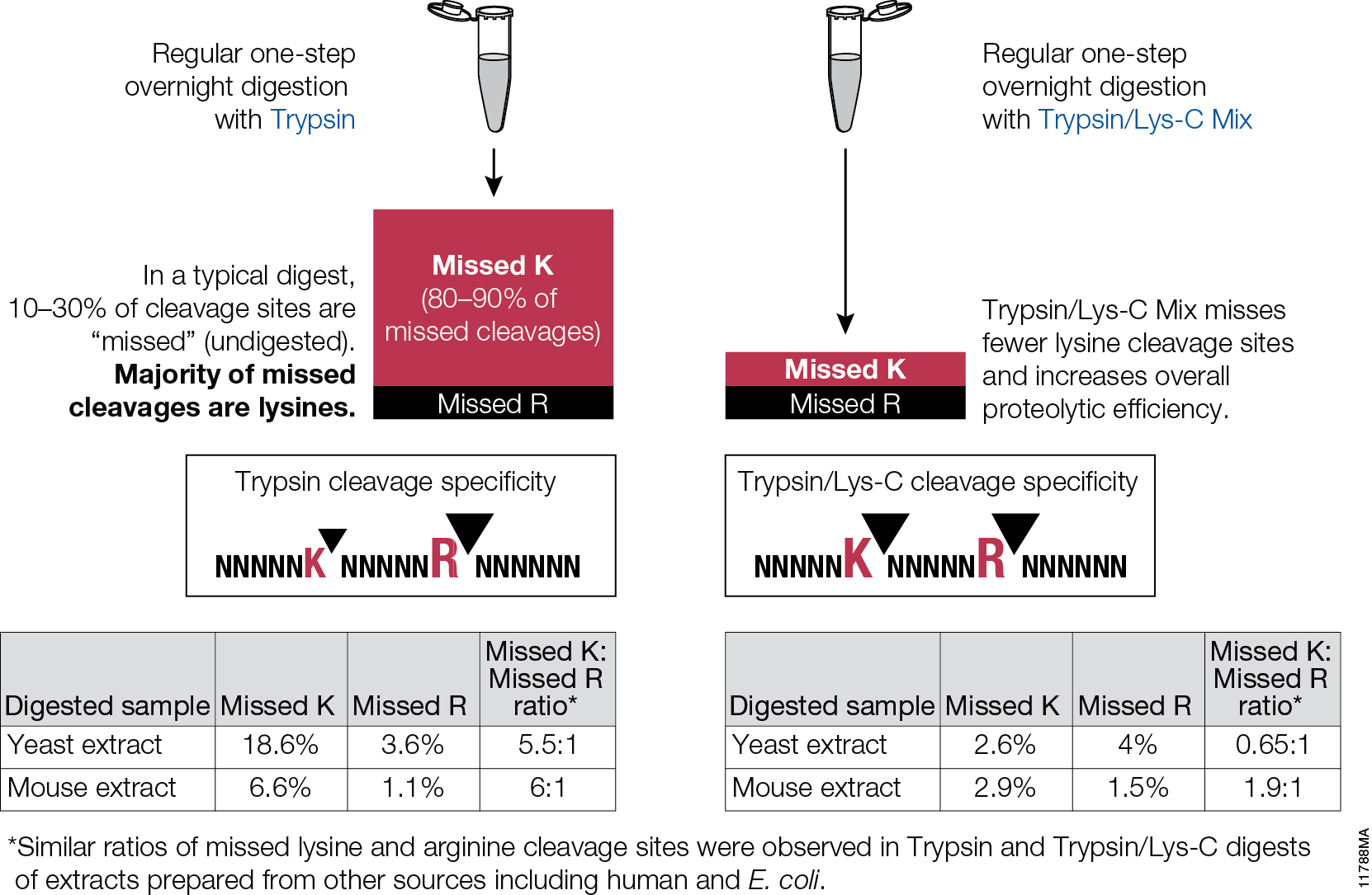
Trypsin/Lys-C Mix, Mass Spec Grade, is a mixture of Trypsin Gold, Mass Spectrometry Grade, and rLys-C, Mass Spec Grade. The Trypsin/Lys-C Mix is designed to improve digestion of proteins or protein mixtures in solution.It is a little known fact that trypsin cleaves at lysine residues with lesser efficiency than at arginine residues. Inefficient proteolysis at lysine residues is the major cause of missed (undigested) cleavages in trypsin digests.

11788MA


Supplementing trypsin with Lys-C enables cleavage at lysines with excepetional efficiency and specificity. Following the conventional trypsin digestion protocol (i.e., overnight incubation at nondenaturing conditions, reduction,alkylation, 25:1 protein:protease ratio [w/w], mix and incubate overnight at 37°C.) Replacing trypsin with Trypsin/Lys-C Mix in this conventional protocol leads to multiple benefits for protein analysis including more accurate mass spectrometry-based protein quantitation and improved protein mass spectrometry analytical reproducibility.

Are you looking for proteases to use in your research?
Explore our portfolio of proteases today.
Site-specific copy number variations in cancer: A story begins to unfold

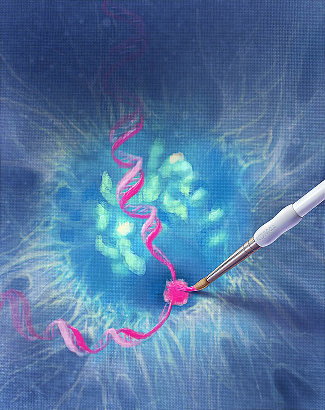
Designed by Nick Klein for ISO-form, courtesy of Promega.

Tumor cells are characterized by many features: including uncontrolled proliferation, to loss of contact inhibition, acquired chromosomal instability and gene copy number changes among them. Some of those copy number changes are site-specific, but very little is known about the mechanisms or proteins involved in creating site-specific copy number changes. In a recently published Cell paper, Black and colleagues, propose a mechanism for site-specific copy number variations involving histone methylation proteins and replication complexes.

Previous work from Klang et al. had shown that local amplification of chromosomal regions occurs during S phase and that chromatin structure plays a critical role in this amplification (2), and other work by Black and colleagues (3) implicated KDM4A in changing timing of replication by altering chromatin accessibility in specific regions. Other research also had shown that KDM4A protein levels influence replication initiation and that KDM4A has a role in some DNA damage response pathways (4,5).  Looking at the body of work, Black et al. hypothesized that KDM4A, with its roles in replication, might possibly provide link into the mechanism of site-specific copy number variation in cancer. Continue reading “Site-specific copy number variations in cancer: A story begins to unfold”

Convenient, Non-Radioactive Detection of Isoaspartate

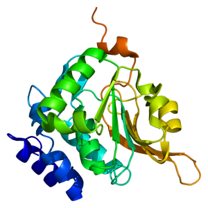
Structure of the PCMT1 protein. Based on PyMOL rendering of PDB 1i1n. Licensed under creative commons http://creativecommons.org/licenses/by-sa/3.0/deed.en

The ISOQUANT® Isoaspartate Detection Kit is intended for quantitative detection of isoaspartic acid residues in proteins and peptides, which can result from the gradual, nonenzymatic deamidation of asparagine or rearrangement of aspartic acid residues.

The ISOQUANT® Kit is designed to provide information regarding the global formation of isoaspartic acid residues at Asn and Asp sites, not at each site separately.

The deamidation of asparagine residues and rearrangement of aspartic acid residues is characterized by the formation of a succinimide intermediate that resolves to form a mixture of isoaspartic acid (typically 70–85%) and aspartic acid.
The rate and extent of isoaspartic acid formation can vary widely among proteins, depending on the amino acid sequence and size of the target protein. Deamidation of Asn residues has been observed most frequently at Asn-Gly and Asn-Ser sites within proteins.

The ISOQUANT® Isoaspartate Detection Kit uses the enzyme Protein Isoaspartyl ethyltransferase (PIMT) to specifically detect the presence of isoaspartic acid residues in a target protein. PIMT catalyzes the transfer of a methyl group from S-adenosyl-L-methionine (SAM) to isoaspartic acid. Spontaneous decomposition of this methylated intermediate results in the release of methanol and reformation of the succinimide.

References:

Wang, W. et al. (2012) Quantification and characterization of antibody deamidation by peptide mapping with mass spectrometry. Int. J. Mass. Spec. 312, 107–13.

Grappin, P. et al. (2011) New proteomic developments to analyze protein isomerization and their biological significance in plants. J. Proteomics, 74, 1475–82.

Yang, H. and Zubarev, R.A. (2010) Mass spectrometric analysis of asparagine deamidation and aspartate isomerization in polypeptides. Electrophoresis 31, 1764–71.

Sinha, S. et al. (2009) Effect of protein structure on deamidation rate in the Fc fragment of an IgG1 monoclonal antibody. Protein Sci. 18, 1573–84.

ProteaseMAX Surfactant: Enhanced In-solution Digestion Applications

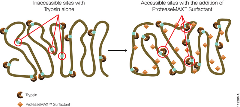
ProteaseMax 11228MA

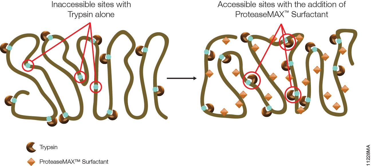
The primary advantage of ProteaseMAX™ Surfactant is that it improves identification of proteins in gel by enhanced protein digestion, increased peptide extraction, and minimized post digestion peptide loss. However, ProteaseMAX™ Surfactant can also facilitate in-solution digestion protocols.

ProteaseMAX™ Surfactant offers two major benefits for digesting proteins in solution.

Continue reading “ProteaseMAX Surfactant: Enhanced In-solution Digestion Applications”

Basic Biology Matters

crop image2Every scientific paper is the story of a journey from an initial hypothesis to a final conclusion. It may take months or years and consists of many steps taken carefully one at a time. The experiments are repeated, the controls verified, the negative and positive results analyzed until the story finally makes sense. Sometimes the end of the story confirms the hypothesis, sometimes it is a surprise. A paper published last week in Cell describes a study where a team of researchers investigating one problem in basic biology (how one component of a signaling complex works), found an unexpected and potentially significant application in a different field (cancer research).

The paper, published in the June 6 issue of Cell, describes a previously unknown interaction between two cellular proteins—the transcription factor HIF1A and the cyclin-dependent kinase CDK8—in the regulation of genes associated with cellular survival under low-oxygen conditions. An accompanying press release describes how the discovery of a role for CDK8 in this process may have implications for cancer research, as CDK8 may be a potential target for drugs to combat “hypoxic” tumors. Continue reading “Basic Biology Matters”

Rabbit Reticulocyte Lysate Translation Systems: Tools for the analysis of translational regulation

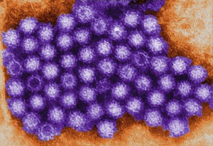
TEM of Norovirus particles. Photo Credit: Charles D. Humphrey, Centers for Disease Control and Prevention

Rabbit Reticulocyte Lysate Translation Systems are used in the identification of mRNA species, the characterization of their protein products and the investigation of transcriptional and translational control. Rabbit Reticulocyte Lysate is prepared from New Zealand white rabbits. After the reticulocytes are lysed, the extract is treated with micrococcal nuclease to destroy endogenous mRNA and thus reduce background translation to a minimum.

Untreated Lysate is prepared from New Zealand white rabbits in the same manner as treated lysates with the exception that it is not treated with micrococcal nuclease. Unlike a coupled system that initiates transcription/translation from DNA, the RNA-based rabbit reticulocyte can be used for the direct investigation of transcriptional/translational control and the replication of RNA-based viruses.


References

Characterization of translation regulation (i.e., UTRs, Capping, IRES)

  1. Nguyen, H-L .et al. (2013) Expression of a novel mRNA transcript for human microsomal epoxide hydrolase is regulated by short reading frames within it 5’ –untranslated region. RNA. 19, 752–66.
  2. Wei, J. et al. (2013) The stringency of start codon selection in the filamentous fungus Neurospora crass. J. Biol. Chem. 288, 9549–62.
  3. Paek Ki-Y. et al. (2012) Cap-Dependent translation without base-by-base scanning of an messenger ribonucleic acid. Nucl. Acid. Res. 40, 7541–51.
  4. Se, and NH. Su.W. et al. (2011) Translation, stability, and resistance to decapping of mRNA containing caps substituted in the triphosphate with BH3. RNA 17, 978–88.
  5. Anderson, D. et al. (2011) Nucleoside modifications in RNA limit activation of 2’-5’ oligoadenylate synthetase and increase resistance to cleavage by RNase L. Nucl. Acid. Res. 39, 9329-38.

RNA virus Characterization

  1. Vashist, S. et al. (2012) Identification of RNA-protein interaction networks involved in the Norovirus life cycle. J. Vir. 86, 11977–90.
  2. Soto-Rifo, R. et al. (2012) Different effects of the TAR structure on HIV-1 and HIV-2 genomics RNA translation. Nucl. Acids. Res. 40, 2653–67.
  3. Poyry, T. et al. (2011) Mechanisms governing the selection of translation initiation sites on Foot-and-Mouth Disease Virus RNA. J.Vir. 85, 10178–88.
  4. Cheng, E. et al. (2011) Characterization of the interaction between Hantavirus nucleopcapsid protein and ribosomal protein S19. J. Biol. Chem. 286, 11814–24.
  5. Vera-Otarola, J. et al. (2011) The Andes Hantavirus NSs Protein is expressed from the Viral mRMA by a leaky scanning mechanism. J. Vir. 86, 2176–87.