Fixed in the Past, Focus on the Future

“I would do more with my samples, but it’s just not possible…I know there’s probably a wealth of information in there, but there is just no way to get it out…I’ve got blocks of tissue sitting in the lab, experiments I want to run, but no good way to get clean nucleic acids out.”

These are a few of the comments I heard when talking with scientists at the American Society of Human Genetics meeting last week in Montreal. They, and countless other researchers, are sitting on a treasure trove of information that may have been locked away a few months ago, a few years ago, or decades ago. I’m referring to formalin-fixed, paraffin-embedded (FFPE) tissue blocks. It is estimated that there are upwards of 400 million tissue blocks archived globally and scientists are clamoring to find ways to best utilize nucleic acids derived from these tissues in applications like qPCR, microarrays, and next generation sequencing.1  Continue reading “Fixed in the Past, Focus on the Future”

Screening for Drug-Drug Interactions with PXR and CYP450 3A4 Activation

The pregnane X receptor (PXR) is a nuclear receptor known to regulate expression of cytochrome P450 (CYP450) drug-metabolizing enzymes (1). PXR has even been designated the “master xenosensor” due to its ability to upregulate cellular levels of a variety of drug-metabolizing enzymes in response to drugs and foreign chemicals. Elevated levels of CYP450 enzymes can elicit alterations in the pharmacokinetics of co-administered drugs, which can result in adverse drug-drug interactions (DDI) or diminished bioavailability. By assessing PXR activation and CYP450 enzyme induction early in the drug development process, many companies hope to reduce late-stage clinical failures and minimize the high costs associated with bringing a new drug to market.

Proportion of drugs metabolized by different CYPs

A paper by Shukla et al. (2) examined over 2,800 clinically used drugs for their ability to activate human PXR (hPXR) and rat PXR (rPXR), induce human cytochrome P450 3A4 enzyme (CYP3A4) at the cellular level, and bind hPXR at the protein level. Several studies have identified PXR as playing a key role in regulating the expression of CYP3A4, an enzyme involved in the metabolism of more than 50% of all drugs prescribed in humans. Since PXR activation and CYP3A4 induction have an impact on drug metabolism and pharmacokinetics, the authors wanted to obtain data that would be valuable in understanding structure-activity relationships (SARs), the connection between chemical structure and biological activity, when prioritizing new molecular entities (NMEs) for further in vitro and in vivo studies.

Continue reading “Screening for Drug-Drug Interactions with PXR and CYP450 3A4 Activation”

Cell-Free Kinase Assays

Protein phosphorylation is one of the most biologically relevant modifications and is involved in many eukaryotic and prokaryotic cellular signaling processes. It is estimated that one-third of human proteins are phosphorylated.

The following examples utilize the ability of cell free experession to express active proteins, and when supplemented with the necessary components (e.g., ATP, NaCl), to be used for the characterization of phosphorylation events.

Modrof, J. et al. (2005) Phosphorylation of bluetongue virus nonstructural protein 2 is essential for formation of viral inclusion bodies. J. Vir. 79, 10023–31. Use of TNT® cell-free to express NS2 and NS2 mutant proteins for use in vitro kinase assays to confirm phosphorylation by protein kinase CK2.

Kwon, S. et al. (2005) Signal pathway of hypoxia-inducible factor-1alpha phosphorylation and its interaction with von Hippel-Lindau tumor suppressor protein during ischemia in MiaPaCa-2 pancreatic cancer cells. Clin. Cancer Res. 11, 7607–13. The TNT® system was used to identify which p38 mitogen-activated protein kinase isoform(s) was cabable of phosphorylation of HIF—1 alpha

Harris, J. et al. (2006). Nuclear accumulation of cRel following C-terminal phosphorylation by TBK1/IKK epsilon. J. Immunol. 177, 2527–35. IKK and IKK mutants were expressed using TNT and used in a vitro kinase assay to characterize the recognition motif in cRel transcription domain

Jailais, Y. et al. (2011) Tyrosine phosphorylation controls brassinosteroid receptor activation by triggering membrane release of its kinase inhibitor. Genes Dev. 25, 232–37. Using a vitro kinase assay, full–length and truncations versions of the Brassinostediod-insentive receptor protein were expressed using the TNT® system and incubated with purified BR11 kinase domain to determine binding sites of the two proteins.
A Scalable and Sensitive Assay for HDAC Activity

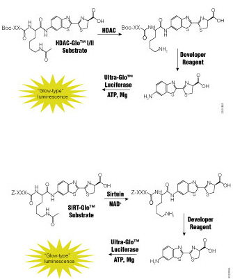
Dysfunction of histone deacetylases (HDACs) is associated with many diseases including cancers, asthma and allergies, inflammatory diseases and disorders affecting the central nervous system. Because of their involvement in such a wide range of pathologies, HDACs have become a target for drug discovery. Traditional HDAC activity assays are either isotopic or fluorescent assays using artificial substrates that are prone to artifacts or fluorescence interference. There is a need for a functional assay that is sensitive, accurate and amenable to drug-screening activities.

A recent paper by Halley et al. in the Journal of Biomolecular Screening describes the evaluation of a bioluminogenic HDAC assay, the HDAC-Glo™ I/II Assays,

Continue reading “A Scalable and Sensitive Assay for HDAC Activity”

Optimized Protein Expression: Flexi Rabbit Reticulocyte Lysate

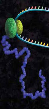
A protein chain being produced from a ribosome.

mRNAs commonly exhibit differing salt requirements for optimal translation. Small variations in salt concentration can lead to dramatic differences in translation efficiency. The Flexi® Rabbit Reticulocyte Lysate System allows translation reactions to be optimized for a wide range of parameters, including
Mg2+ and K+ concentrations and the choice of adding DTT. To help optimize Mg2+ for a specific message, the endogenous Mg2+ concentration of each lysate batch is stated in the product information included with this product.

The following references utilize the features of Flexi Rabbit Reticulocyte Lysate System to investigate certain parameters of translation:

Vallejos, M. et al. (2010)The 5′-untranslated region of the mouse mammary tumor virus mRNA exhibits cap-independent translation initiation. Nucl Acids Res. 38, 618–32. Identification of internal ribosomal ribosomal entry site in the 5’ untranslated region of the mouse mammary tumor virus mRNA.

Spriggs, K. et al. (2009) The human insulin receptor mRNA contains a functional internal ribosome entry segment. Nucl. Acids. Res. 17, 5881–93. Identification of a functional internal ribosome entry site in the human insulin receptor mRNA.

Powell, M. et al. (2008) Characterization of the termination-reinitiation strategy employed in the expression of influenza B virus BM2 protein. RNA 14, 2394–06. Analysis of the mRNA signals involved in the expression of influenza B virus BM2 protein.

Sato, V. et al. (2007) Measles virus N protein inhibits host translation by binding to eIF3-p40. J. Vir. 81, 11569–76. Charaterized the effect of the measles virus N protein binding to the translation initiation factor eIF3-p40 on the expression of various proteins in rabbit reticulocyte lysate.

Hirao, K. et al. (2006) EDEM3, a soluble EDEM homolog, enhances glycoprotein endoplasmic reticulum-associated degradation and mannose trimming. J. Biol. Chem. 281, 9650–58. The EDEM3 protein was expressed in the presence of canine microsomal membranes to establish that co-translational translocation occurs into the endoplasmic reticulum.

Shenvi, C. et al. (2005) Accessibility of 18S rRNA in human 40S subunits and 80S ribosomes at physiological magnesium ion concentrations–implications for the study of ribosome dynamics. RNA 11, 1898–08. Characterization of ribosome dynamics under different ionic conditions.

Nair, A. et al. (2005) Regulation of luteinizing hormone receptor expression: evidence of translational suppression in vitro by a hormonally regulated mRNA-binding protein and its endogenous association with luteinizing hormone receptor mRNA in the ovary. J. Biol. Chem. 280, 42809–16. Examined the affect of luteinizing hormone receptor mRNA binding protein on transltional suppression of luteinizing hormone receptor RNA.

Optimization of Western Blots Detecting Proteins Synthesized Using Cell-Free Expression #2

Detection of protein expressed using cell-free systems is required for most applications such as protein:protein interaction and protein:nucleic acid interaction studies. Traditionally, one adds radioactive [35S]methionine to cell-free expression reactions, and the methionine is incorporated into the expressed protein, allowing detection by autoradiography. Many researchers are moving away from radioactivity. Traditional Western blot analysis provides the researcher a nonradioactive method for detection but, if performed improperly, can result in high background, which can mask expressed proteins and affect downstream applications.

One critical step in producing low-background, high-signal Western blots is choosing the correct dilution of the primary antibody. Typically the manufacturer recommends antibody dilution from 1:1,000 to 1:2,500 for standard western blotting experiments. However when using crude lysates as a source of the target protein, these recommendations exhibit significant background. When the antibody was diluted 1:50,000, background was decreased significantly, and the positive signal was a large percentage of the total signal.

As a general recommendation when performing Western blot analysis of proteins expressed in cell-free systems, one must experimentally determine the optimal dilution of the primary antibody. In the Western blots performed in this study, primary antibodies were diluted ~50-fold more than the provider’s recommended dilution.

For additional technical details refer to this recent article published in Promega’s PubHub:

Hook, B and Schagat, T. (2011) Non-Radioactive Detection of Proteins Expressed in Cell-Free Expression Systems Promega Corporation Web site. Accessed August 17, 2011.

A New Method that Marks Proteins for Destruction

The ability to manipulate genes and proteins and observe the effects of specific changes is a foundational aspect of molecular biology. From the first site-directed mutagenesis systems to the development of knockout mice and RNA interference, technologies for making targeted changes to specific proteins to eliminate their expression or alter their function have made tremendous contributions to scientific discovery.
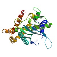
A recent paper highlights a novel application of HaloTag technology to enable the targeted destruction of specific HaloTag fusion proteins in vivo. The paper, published online in the July issue of Nature Chemical Biology, details a promising new method with application for validation of potential drug targets by specific in vivo inhibition, and for studying the function of specific genes in organogenesis or disease development. Continue reading “A New Method that Marks Proteins for Destruction”

Identifying and Profiling Inhibitors for PI 4-Kinases Using a Luminescent High-Throughput Screen

The phosphotidylinositol 4-kinases (PI 4-kinases) generate phosphotidyl-4-phosphate (PI(4)P) from phosphotylinositol. PI(4)P is an important precursor for other phosphoinositides involved in signaling, such as PI(4,5)P2, which is the substrate of phospholipase C (PLC) and the precursor of DAG and insitol (1,4,5) triphosphate.

There are four different mammalian PI 4-kinases currently described, and these have been divided into two classes based on their sensitivities to wortmannin and adenosine. Type II PI 4-kinases (PI4K2A and PI4K2B) are not sensitive to wortmannin, but are inhibited by the nonspecific inhibitor adenosine; Type III PI 4-kinases (PI4KA and PI4KB) are sensitive to wortmannin.

The functions of the PI 4-kinases and their products are not fully understood. At least one study has shown that PI 4-kinases are important for the proper recycling of synaptic vesicles. The PI 4-kinase from Drosophila, four-wheel drive, is critical for contractile ring formation during cytokinesis. Other studies in yeasts and mammals have shown that PI 4-kinases localize to the Golgi, and in mammals might be critical for proper budding of vesicles from the Golgi. Additionally, type III PI 4-kinases appear to play a role in the replication of hepatitis C virus (HCV) and enteroviruses by participating in the formation of altered host membrane structures. Although, we have hints about their function, to really understand and dissect the precise roles of PI 4-kinases in cells, new tools, such as specific small-molecule inhibitors are required.

Continue reading “Identifying and Profiling Inhibitors for PI 4-Kinases Using a Luminescent High-Throughput Screen”

Troubleshooting T-Vector Cloning

Why do few of my pGEM®-T or pGEM®-T Easy Vector clones contain the PCR product of interest?

There are several possible reasons why the PCR product may not be recovered after ligation, bacterial transformation and plating when using the pGEM®-T or pGEM®-T Easy Vector Systems.

The PCR fragment may not be A-tailed. Without the A overhangs, the PCR product cannot be ligated into a T vector. Use a nonproofreading DNA polymerase like GoTaq® DNA Polymerase for PCR. If a proofreading DNA polymerase is used, A overhangs will need to be added. Purify the PCR fragment, and set up an A-tailing reaction (see the pGEM®-T and pGEM®-T Easy Vector Systems Technical Manual #TM042). The A-tailed product can be added directly to the ligation as described in the pGEM®-T or pGEM®-T Easy Vector protocol.

The insert:vector ratio may not be optimal. The ideal ratio for each insert to a vector can vary. For example, the Control Insert DNA works well at a 1:1 ratio, but another insert may be ligated more efficiently at a 3:1 ratio. Check the integrity and quantity of your PCR fragment by gel analysis. Optimize the insert:vector ratio (see Technical Manual #TM042).

Multiple PCR products were amplified and cloned into the pGEM®-T or pGEM®-T Easy Vector. Other amplification products including primer dimers will compete for ligation into the T vector, decreasing the possibility that the desired insert will be cloned. To minimize other competing products, gel purify the PCR fragment of interest.

Promega Technical Services Scientists are here to assist you in troubleshooting your experiments at any time. Contact Technical Services.

Related Posts

Cell-Free Applications: RNA Toeprinting

A protein chain being produced from a ribosome.
Precise mapping of the positions of ribosomes and associated factors on mRNAs is essential for characterizing the mechanism of translation. Using the toeprinting assay, mRNA is translated using purified components or crude cell lysates such as rabbit reticulocyte. Cycloheximide is added to the reaction to inhibit elongation. This arrests the position of the ribosomes on the mRNA transcript. The mRNA/ribosomal complex are then copied into cDNA by reverse transcriptase using a complementary radiolabeled primer. Where the reverse transcriptase meets the ribosome bound to the mRNA, cDNA extension is halted, and a toeprint cDNA fragment is generated.

The following references use rabbit reticulocyte lysates as the basis for toeprinting experiments to better understand the mechanism of translation.

Weill, L. et al. (2010)Nucl. Acid, Res. 38, 1367–81. A combination of chemical/enzymatic analyses indicated that gag open reading frame of three viruses adopts a stable secondary structure that allows IRES mediated translation. Mutations that destabilized conserved elements severely inhibit translation. Additional analysis via toeprinting showed HIV-2 IRES has the unique ability to attract up to three initiation complexes on a single RNA molecule.

De Breyne, S. et al. (2008) RNA 14, 367–80. The Simian picornavirus type 9 (SPV9) genome contains a group of IRES that resembles hepacivirus/pestvirus (HP) IRES. Characterization of the initiation process using the toeprinting assay in correlation with other techniques revealed aspects that resemble initiation on the HP IRES and others that are unique to SPV9.

Andreev, D. et al. (2008) RNA 14, 233–39. Rel E is a well characterized toxin involved in the nutritional stress response in bacteria and archae. Rel lacks any eukaryote homolog. Based on toeprinting data, it was demonstrated that RelE cleaves mRNA in the A site of the eukaryote ribosome.