Riboprobes: RNA Probes Are Still Valuable Research Tools

9613ca[1]Riboprobes are RNA probes that can be produced by in vitro transcription of cloned DNA inserted in a suitable plasmid downstream of a viral promoter.
Viruses code for their own RNA polymerases, which are highly specific for the viral promoters. Using these enzymes, labeled NTPs, and inserts in both forward and reverse orientations, both sense and antisense riboprobes can be generated from a cloned gene.
Transcription of RNA is performed with the appropriate RNA polymerase (T3, T7 or SP6), depending on the RNA polymerase promoter sites present in the chosen vector. Because these polymerases are extremely promoter-specific (i.e., there is almost no transcriptional cross talk), virtually homogeneous RNA can be obtained using plasmid DNA as the template in a transcription reaction. When it is desirable to copy only insert DNA sequences, the plasmid is linearized at an appropriate restriction site before the transcription reaction and only discrete “run-off” transcripts are obtained, virtually free of vector sequences. RNA transcripts may be used to generate radioactive probes for hybridization to Northern and Southern blots, plaque and colony lifts as well as non-radioactive probes (i.e, labeled with digoxgenin)for in situ hybridization.

Recent references using riboprobes include: Continue reading “Riboprobes: RNA Probes Are Still Valuable Research Tools”

RNAi: The Dream Makes a Comeback

This Promega Notes Cover from 2004 celebrated the potential stop and go power of DNA-directed RNAi.

In the early 2000s, RNAi was a hot topic. The science world was abuzz with all the possibilities that harnessing this natural process could hold. And why not? The idea of posttranscriptionally silencing genes using only a small fragment of double-stranded RNA is huge—big enough to earn the scientists who discovered it a Nobel Prize in 2006.

The process of RNAi starts with short (~70 nucleotieds), double-stranded fragments of RNA called short hairpin RNAs (shRNA). These shRNAs are exported into the cytoplasm and cleaved by the enzyme Dicer into smaller pieces of RNA that are about 21 nucleotides long and are referred to as small interfering RNAs (siRNA). The siRNAs reduce or stop expression of proteins through a sequence of events where the antisense strand of the siRNA is incorporated into and RNA-induced silencing complex (RISC), which then attaches to and degrades its complimentary messenger RNA, thereby reducing or completely stopping expression.

It turned out, however, that harnessing the promise of RNAi was a little trickier than anticipated. Continue reading “RNAi: The Dream Makes a Comeback”

Promising Treatment for Marburg Virus Hemorrhagic Fever

I admit to some trepidation about the diseases that may be harbored in my backyard. For example, do the mice in my yard and, despite my and my cats’ efforts, in my house carry deer ticks that harbor the bacterium Borrelia burgdorferi, which causes Lyme disease? Should I be keeping an eye on the vitality of the birds around my property and density of my local mosquito population for potential risk of West Nile Virus transmission? As troublesome as these infections can be, mortality is low for infected humans. Contrast that with the mortality rate of up to 90% for the filoviruses Ebola and Marburg. I find it easy to dismiss these viruses because the reservoir (asymptomatic host) is not in the Upper Midwest but rather Africa, but the tragedy of the Ebola outbreak in the West African countries of Liberia, Sierra Leone and Guinea demonstrates the number of lives lost in an epidemic. Currently, there is no therapy or vaccine to treat these deadly viruses other than transferring antibodies from survivors to those infected. Therefore, the article in Science Translational Medicine about an antiviral treatment that protected macaques injected with a lethal dose of Marburg virus was welcome news.

Continue reading “Promising Treatment for Marburg Virus Hemorrhagic Fever”

Chikungunya Virus and the Promise of a Virus-Like Particle Vaccine

My family and I just returned from a week-long camping trip along the North Shore of Lake Superior in Minnesota. It is beautiful country, filled with lakes, rivers, ponds—and mosquitoes, lots and lots of mosquitoes. We went prepared for the worse. We had a screen tent, head nets and tubes and tubes of insect repellent because in this area of the world, mosquitoes are a flying, buzzing, picnic-ruining, itch-inducing pest. In the US, though, a pest is really all they are. In other areas of the world they are a flying, buzzing, disease-carrying, deadly menace.

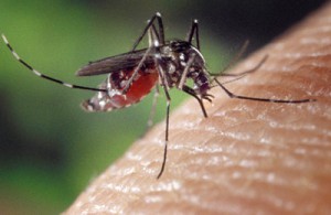
Image courtesy of James Gathany and the CDC

Mosquitos act as vectors for many diseases including malaria, Dengue fever, Yellow fever, encephalitis, West Nile Virus and chikungunya virus. Many of these diseases are deadly; in fact, mosquitoes are responsible for more human deaths than any other animal (~725,000 deaths annually). Although most of these diseases have a long and infamous history, two of them, West Nile virus (first identified in 1932) and chikungunya virus (first identified in 1950), are relative new comers on the world health stage. Continue reading “Chikungunya Virus and the Promise of a Virus-Like Particle Vaccine”

Novel Cell Surface Markers Identified that Differentiate White, Beige and Brown Adipocytes

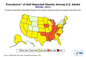
2012 CDC-based data on U.S. obesity percentages by state.

Estimates of obesity in the U.S. range from 30% (Centers for Disease Control data) to 70% (persons selling online and television audience-focused weight-loss programs). We are a nation of fat or fat-obsessed persons, and rightfully so. CDC data shows that the cost of obesity, in 2008 dollars, was estimated at $147 billion. That amount of money would buy a lot of french fries or cheesecake or __ (name your poison).

We all help pay those high-dollar amounts in terms of rising healthcare costs, thus there is considerable interest in finding ways to not only avoid, but also to combat obesity.

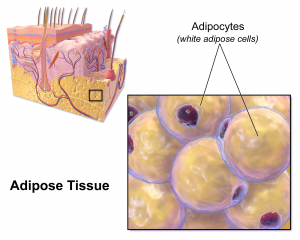
In recent years researchers working to understand body fat biology have produced exciting information on differences in types of fat. For instance, we now understand that in addition to white adipose tissue, animals and humans also have brown and beige adipose tissue. White adipose tissue or WAT is commonly found in humans and mice subcutaneously and in visceral fat. Brown adipose tissue or BAT, and beige adipose, is less common, and in humans and mice, is found in deeper cervical, supraclavical and paraspinal areas.

Continue reading “Novel Cell Surface Markers Identified that Differentiate White, Beige and Brown Adipocytes”

Angel’s Glow: Bioluminescence Uncovered on the Battlefield

New information has surfaced about this story, and we encourage you to read our updated blog from July 2024 (linked) for the latest on this story.

1888 Chromolithograph of the Battle of Shiloh, American Civil War, produced by L. Prang & Co.

If battlegrounds could speak they would have many stories to tell.  In some cases the microbes found in those soils have lived on to separate fact from fiction. One such story has its origins in the Battle of Shiloh, which went down in history as one of the bloodiest battles fought during the American Civil War.  As the soldiers lay mortally wounded on the cold, hard grounds of Shiloh waiting for medical aid, they noticed a very strange phenomenon. Some of the wounds actually appeared to be glowing in the dark casting a faint light into the darkness of the battlefield. And the legend goes that soldiers with the glowing wounds had a better chance at survival and recovery from infections than their fellow brothers-in-arms whose wounds were not similarly luminescent. The seemingly protective effect of the mysterious light earned it the moniker “Angel’s Glow.”

Fast forward to the 21st century.

Continue reading “Angel’s Glow: Bioluminescence Uncovered on the Battlefield”

Hope for Treatment of Carbapenem-Resistant Bacteria

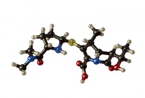
Structure of the antibiotic meropenem
Last month brought some hopeful news on the subject of antibiotic resistance. A paper published in Nature on June 26 described the isolation of a fungal compound that restored the antibiotic sensitivity of carbapenem-resistant enterobacteria. An editorial accompanying the paper took encouragement from the article–considering it a sign that the well of potential sources of new antimicrobial agents, and agents that inhibit resistance mechanisms, is not yet dry:

But the reservoir of natural products with the potential to act as antibacterial drugs has not yet been exhausted. In contrast to general thinking by drug companies, screening for such products may well still have a bright future” Nature News and Views: “Antibiotic resistance: To the rescue of old drugs” Meziane-Cherif & Courvalin, Nature 510, 477–478.

The emergence of bacteria that are resistant to antibiotics has been an object lesson in the relentlessness of natural selection; the moment a new antibiotic is developed and introduced, the countdown to the emergence of resistance begins. The race to keep the one step ahead of emerging resistance mechanisms has been going on since antibiotics were first introduced.

The history of the development of penicillin and related antibiotics is both an illustration of the ingenuity of scientists and of the never-ending nature of this battle with emerging resistance. The Nature paper is the latest installment in that story. Continue reading “Hope for Treatment of Carbapenem-Resistant Bacteria”

Screening for Antiviral Compounds under Level 4 Containment Conditions

Working with bacteria and viruses that cause life-threatening diseases with no currently available treatment options takes guts. Most scientists are familiar with the routine requirements of good aseptic technique, are highly aware of laboratory safety requirements, and are more than familiar with autoclaves and sterilization issues, but if we make a mistake the consequences are usually only lost time or a spoiled experiment—not a lost life.

Scientists working with highly virulent organisms deal with a whole other level of risk that requires adherence to the strictest of safety regulations, and these containment regulations can sometimes place constraints on the type of experiment that can be performed with dangerous pathogens. A paper published in the April 2014 issue of Assay and Drug Development Technologies brought this to my attention and reminded me of the serious issues some scientists face on a daily basis as they research ways to combat infectious diseases.

Continue reading “Screening for Antiviral Compounds under Level 4 Containment Conditions”