Weaving Tangled Webs with Cell-Free DNA

The ability to isolate and assay circulating cell-free DNA from plasma holds promise for improved diagnostics and treatment in the clinic. The use of blood-based non-invasive prenatal testing (NIPT) has been well described. Such testing is based on circulating cell-free fetal DNA in blood of a pregnant woman for diagnosis and screening  of chromosomal anueploidy (e.g. Trisomy 21, Down Syndrome), sex-linked diseases, and genetic diseases that are known to result from a specific mutation in a single gene (1). Additionally, most cancers carry somatic mutations that are unique to the tumors, and dying tumor cells release small pieces of their DNA into the blood stream (2). This circulating cell-free tumor DNA can be used as a biomarker to “follow” cancer progression or regression during treatment, and diagnostic methods also are being developed to detect even early stage cancers from circulating tumor DNA (3). Further, increases in circulating cell-free DNA have been well documented after intense exercise, trauma, sepsis and even associated with autoimmune diseases such as system lupus erythematosus (SLE; 1,4). In these latter examples increases in extracellular DNA are associated with evolutionarily conserved innate immune responses involving the production of neutrophil extracellular traps (NETs). Monitoring the circulating cell-free DNA of NETs has implications for treatment and diagnosis of autoimmune diseases, cardiovascular events and traumatic injuries (4–7).

How Neutrophils Weave a Defensive Web

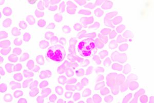
Blood smear showing two prominent neutrophils in the field of view

Neutrophils are the most abundant type of white blood cell and are part of the innate immune response, participating in non-specific immune responses to injury or pathogens. They are one of three types of granuolcytes, and can be recognized by their multi-lobed nucleus and the prominent granules that fill their cytoplasm. Generally they are first to the scene of injury or infection. Early in my scientific career, I was taught that neutrophils fought disease via phagocytosis and occasionally by firing a barrage of toxic enzymes and molecules at invaders. Mostly though they released cytokines that recruited the “important” cells of the specific immune system to the area.

For these reasons, I never really thought much about neutrophils. That is until recently, when I learned about Neutrophil Extracellular Traps (NETs). It turns out that neutrophils are pretty awesome, sacrificing themselves in a cloud-like explosion of DNA, chromatin, and granule proteins

Continue reading “Weaving Tangled Webs with Cell-Free DNA”

Of Elephant Research and Wildlife Crime – Molecular Tools that Matter

Here at Promega we receive some interesting requests…

Take the case of Virginia Riddle Pearson, elephant scientist. Three years ago we received an email from Pearson requesting a donation of GoTaq G2 Taq polymerase to take with her to Africa for her field work on elephant herpesvirus. Working out of her portable field lab (a tent) in South Africa and Botswana, she needed a polymerase she could count on to perform reliably after being transported for several days (on her lap) at room temperature. Through the joint effort of her regional sales representative in New Jersey/Pennsylvania (Pearson’s lab was based out of Princeton University at the time) and our Genomics product marketing team, she received the G2 Taq she needed to take to Africa. There she was able to conduct her experiments, leading to productive results and the opportunity to continue pursuing her work.

Continue reading “Of Elephant Research and Wildlife Crime – Molecular Tools that Matter”

The Birth of a Disease? A New Anthrax-Like Disease Found in Sub-Saharan Africa

Copyright
Copyright

It usually starts with one; one dead animal, one sick individual, one case that a doctor thinks is unusual. These are all ways that a new disease makes its presence known. In the case of Bacillus cereus biovar anthracis, it started with a dead chimpanzee (1).  

The wild western chimpanzee was found dead in Côte d’Ivoire in 2001. An investigation led by scientists from the Robert Koch Institute in Berlin identified the pathogenic cause of death to be an atypical B. cereus isolate that caused an anthrax-like disease. Continue reading “The Birth of a Disease? A New Anthrax-Like Disease Found in Sub-Saharan Africa”

Digging Up More Clues in the History of the Black Death

Bubonic plague victims in a mass grave in 18th century France. By S. Tzortzis [Public domain], via Wikimedia Commons
My last blog post on the Black Death highlighted research that suggested that the reintroduction of Yersinia pestis, the causative agent of the pandemic, originated in Europe during the 14–18th centuries rather than from Asia, the hypothesized origin. In my post, I wrote about my curiosity regarding what an Asian skeleton positive for Y. pestis from that same time period would reveal about the strain or strains that were circulating. Well, a team of researchers has been exploring the issue of strain circulation and an Asian connection, and recently published what they gleaned from additional historic Y. pestis samples in Cell Host & Microbe.

Teeth from 178 individuals in three different locations (two European, one Asian) were screened for Y. pestis infection using the plasminogen activator (pla) gene. Continue reading “Digging Up More Clues in the History of the Black Death”

Magnetic Bacteria Carry Drugs into Tumors

cancer cell

At first glance, the biology of magnetic, underwater-dwelling, oxygen-averse bacteria may seem of little relevance to our most pressing human health problems. But science is full of surprises. A paper published in Nature Nanotechnology presents an inspired use of these bacteria to deliver anti-cancer drugs to tumors, specifically targeting the oxygen-starved regions generated by aggressively proliferating cells.

Continue reading “Magnetic Bacteria Carry Drugs into Tumors”

An Epizootic for the Ages: Revisiting the White-Nose Syndrome Story

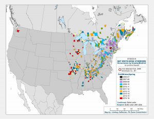
Map showing the spread of WNS across North America

In March 2016, two hikers on a trail east of Seattle, WA, found a little brown bat lying on the ground in obviously poor condition. The bat was taken to an animal shelter where it died two days later from White-Nose Syndrome (WNS).

This bat was the first case of WNS found west of the Rocky Mountains. It represented a jump in the spread of WNS, and a troubling one. WNS was first detected in a cave in Albany, New York, and since then it has been moving slowly westward at a rate of about 200 miles per year, according to David Blehert of the United States Geological Survey, the laboratory that confirmed the WNS diagnosis for the Washington bat. Before this year’s discovery outside of Seattle, the westward-most case detected was in eastern Nebraska.

WNS, caused by a cold-loving fungus, Psuedogymnoascus destructans (Pd), can kill 100% of the hibernating bats in a colony, and in the ten years since it has been detected and monitored has killed over 6 million bats in the United States and Canada. As of July 2016, bats infected with the fungus have been found in 29 states and 5 Canadian provinces.

According to Blehert, this is probably the “most significant epizootic of wildlife” ever observed; never before have we seen hibernating mammals specifically affected by a skin fungus. What does that mean? Are we looking at extinction for some bat species? What are the ecological consequences of rapidly losing so many individuals to disease so quickly? And, what, if anything, can be done to combat the disease and help bat populations recover?

Continue reading “An Epizootic for the Ages: Revisiting the White-Nose Syndrome Story”

Shining Light on a Superbug: Clostridium difficile

Antibiotic-resistant bacteria and their potential to cause epidemics with no viable treatment options have been in the news a lot. These “superbugs,” which have acquired genes giving them resistance to common and so-called “last resort” antibiotics, are a huge concern as effective treatment options dwindle. Less attention has been given to an infection that is not just impervious to antibiotics, but is actually enabled by them.

33553646_l

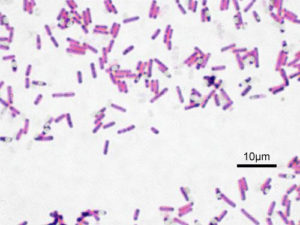
Clostridium difficile Infection (CDI) is one of the most common healthcare-associated infections and a significant global healthcare problem. Clostridium difficile (C. diff), a Gram-positive anaerobic bacterium, is the source of the infection. C. diff spores are very resilient to environmental stressors, such as pH, temperature and even antibiotics, and can be found pretty much everywhere around us, including on most of the food we eat. Ingesting the spores does not usually lead to infection inside the body without also being exposed to antibiotics.

Individuals taking antibiotics are 7-10 times more likely to acquire a CDI. Antibiotics disrupt the normal flora of the intestine, allowing C. diff to compete for resources and flourish. Once exposed to the anaerobic conditions of the human gut, these spores germinate into active cells that embed into the tissue lining the colon. The bacteria are then able to produce the toxins that can cause disease and result in severe damage, or even death.

Continue reading “Shining Light on a Superbug: Clostridium difficile”

Analyzing the Effects of Yersinia pestis Infection on Gene Expression

Yersinia pestis. See page for author [Public domain], via Wikimedia Commons
While scientists using ancient DNA analysis are learning how Yersinia pestis developed over time into the causative agent of three worldwide pandemics, there is still much to learn about the early hours and days of an organism infected with the plague. Y. pestis still infects humans so any insight into disease progression is useful for determining treatment timing and even developing novel treatments to supplement or replace antibiotics. A 2012 study observed how Y. pestis injected into mice spread throughout the body using bioluminescent imaging to track the infection. More recent research reported in PLOS ONE used a more real-world route of infection by introducing an aerosolized Y. pestis to a nonhuman primate model and tracking the transcripts altered during the first 42 hours of infection. Continue reading “Analyzing the Effects of Yersinia pestis Infection on Gene Expression”

Congenital Cytomegalovirus: The Most Common Congenital Infection That You Have Never Heard Of

infant

Down Syndrome. Fetal alcohol syndrome. Spina bifida. Most people have heard of these well know congenital conditions, and know at least in a general sense that they have profound and lasting effects on the children born with them as well as their families. Unfortunately, people are much less aware of a congenital infection that is more common that any of these and affects more infants than all three of the conditions listed above. In fact, this congenital infection causes more cases of congenital disease than all of the 29 conditions currently screened for in most American states combined (1; 2), and yet it is not widely known by the general public. Cytomegalovirus, or CMV, is the most common congenital viral infection in developed countries (3; 4), and the leading cause of congenital sensorineural hearing loss and psychomotor retardation (1).

Continue reading “Congenital Cytomegalovirus: The Most Common Congenital Infection That You Have Never Heard Of”

Zika Virus: Another RNA Virus Emerges

no mosquito

Zika virus has been in the news recently due to growing concerns about its global spread. If you have never heard of Zika virus before, you are not alone. Although first discovered in the 1940s, Zika has not been the subject of much study as infection is considered rare and the symptoms mild. However, all this has changed in recent months due to the rapid spread of the virus in Latin America, where it has been associated with an increased incidence of microcephaly, a severe birth defect where babies are born with underdeveloped brains. Although the connection of Zika with microcephaly is not yet proven, the circumstantial evidence is strong, leading the World Health Organization to declare the spread of Zika virus an international public health emergency earlier this week.

Continue reading “Zika Virus: Another RNA Virus Emerges”