Manipulating Koala Microbiomes Using…Poo Pills?

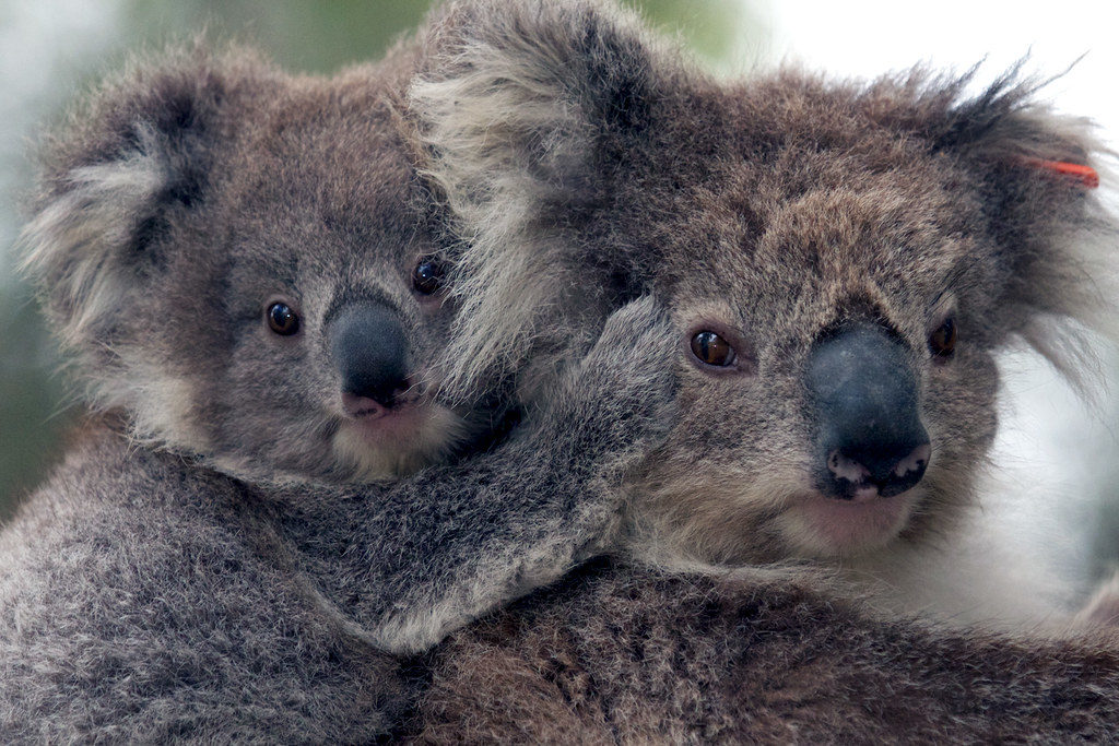
“Koalas – mum and baby” by Amanda Penrose is licensed under CC BY-NC-SA 2.0 

In recent years, it’s become a well-documented fact that koalas are about as picky as they are adorable. These beloved Australian marsupials have evolved to become ecological specialists: consumers that feed primarily on a single organism, or small number of organisms. Eucalyptus, their organism of choice, encompasses approximately 900 species, most of which are native to Australia. To the koala’s benefit, the leaves of eucalyptus trees are difficult to digest, low in protein content and their chemical composition contains compounds that are toxic. This makes their competition for eucalyptus with other species virtually nonexistent.

That’s not to say there isn’t competition amongst themselves. Of those 900 species of eucalyptus, koalas are only really known to feed on about 40–50 of them, and of those 40–50, they tend to limit their diet to around 10. Depending on their location, however, some koalas will only stick to one preferred type, which can lead to trouble.  

Continue reading “Manipulating Koala Microbiomes Using…Poo Pills?”

Delving into the Diversity of The Plague of Justinian

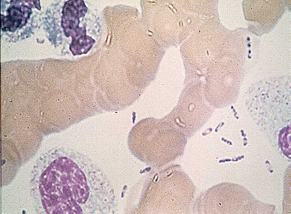
Wayson stain of Yersinia pestis showing the characteristic
Yersinia pestis. U.S. Center for Disease Control [Public domain], via Wikimedia Commons.
Human teeth play a key role in our understanding of how organisms evolve. Whenever a possible new member of the hominid family is uncovered, the shape and number of teeth are used to place that individual in the family tree. Teeth also harbor information about pathogens that have plagued humans for millennia. Because bacteria use our bloodstream as a transport system, protected places that can preserve DNA—like the pulp of teeth—are a rich medium for uncovering information about humans and the microbes that infected them.

Teeth have been the choice for identifying the infectious agent behind the Plague of Justinian in the sixth century and the Black Plague in the 14th century. In fact, Yersinia pestis, the bacterium responsible for these plagues, has infected humans as far back as the Neolithic. But what can we learn about the pandemic strain or strains of Y. pestis described in historical records? A team of researchers from Europe and the US, many of whom have been delving into the history of Y. pestis for the last decade, wanted to further investigate the Plague of Justinian. They studied bacterial DNA extracted from human remains found in Western European communal graves that were dated to around 541–750, the period of the historically documented Plague of Justinian. Their investigation examined the bacteria’s diversity and how far it spread during this “First Pandemic” of plague. Continue reading “Delving into the Diversity of The Plague of Justinian”

Immunotherapy—Don’t Forget the Microbiome

Bacteria make you sick. The idea that bacteria cause illness has become ingrained in modern society, made evident by every sign requiring employees to wash their hands before leaving a restroom and the frequent food recalls resulting from pathogens like E. coli. But a parallel idea has also taken hold. As microbiome research continues to reveal the important role that bacteria play in human health, we’re starting to see the ways that the microbiota of the human body may be as important as our genes or environment.

The story of how our microbiome affects our health continues to get more complex. For example, researchers are now beginning to understand that the composition of bacteria residing in your body can significantly impact the effects of therapeutic drugs. This is a new factor for optimizing drug response, compared to other considerations such as diet, interaction with other drugs, administration time and comorbidity, which have been understood much longer.

Continue reading “Immunotherapy—Don’t Forget the Microbiome”

Research-Based Training for Sustainable Use and Management of Marine Ecosystems in Namibia

In my science blog research/writing, news reports are usually pulled from US sources. But interesting scientific research is obviously being conducted in many places around the globe. When this story from Namibia came along, there was so much I didn’t know. It was time to catch up.

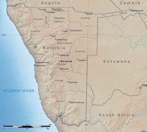
relief map of Namibia
Relief map of Namibia. Image by Natural Earth and Kbh3rd with permission under Wikimedia commons.

Namibia is Exactly Where in Africa?

Namibia is one of the world’s youngest countries, having gained independence from South Africa in 1990. Situated northwest of the country of South Africa on the Atlantic Ocean, Namibia is arid, composed largely of desert.

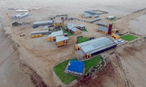
This blog is about research conducted at the Sam Nujoma Research Center, University of Namibia, on Henties Bay. Henties Bay (not shown on this map) is in the region of Erongo, located in the center of Namibia along the coast. Henties Bay has become a tourist destination in part due to the abundance of fish and marine life found there.

Sam Nujoma Research center.
The Sam Nujoma Research Center of University of Namibia, located near Henties Bay.

Continue reading “Research-Based Training for Sustainable Use and Management of Marine Ecosystems in Namibia”

Are We Doing Enough to Stop Candida Auris Infections?

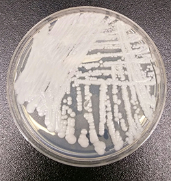
Image of C. auris on plate.
The creamy colonies of C. auris look innocuous. Don’t be fooled. Photo by Shawn Lockhart – Centers for Disease Control, Public Domain, https://commons.wikimedia.org/w/index.php?curid=54680002

Life in the 21st century is full of electronic devices and apps purported to make life easier. Many of us can binge watch movies, videos and news on our phones. There are wireless headphones, electric bicycles, self-operating vacuum cleaners, wine in boxes with taps—and so much more.

This life is, however, not without challenges.

In the event that you or yours ends up in the hospital, the stay could be complicated by an unplanned, unwanted and potentially lethal infection.

No thanks to the yeast, Candida auris.

Continue reading “Are We Doing Enough to Stop Candida Auris Infections?”

Expanding the Plague Family Tree: Yersinia pestis in the Neolithic

Yersinia pestis. See page for author [Public domain], via Wikimedia Commons

In recent years, scientists have been able to refine their molecular tools to resurrect ancient DNA from human graves and determine that yes, Yersinia pestis was the causative agent for the Black Death in the 14th century and the Plague of Justinian in the 6th century.  As more and more human graves have been uncovered, their DNA has revealed many secrets that scientists even ten years ago were unable to discover. With the ability to sequence entire genomes of bacteria that died with their hosts hundreds and even thousands of years ago, researchers are exploring the rise and possible spread of Y. pestis. Each new member sequence adds to the Y. pestis family tree, pinpointing the origin of this bacteria as it diverged from its ancestor Y. pseudotuberculosis. Peering into the past, scientists have been able to track down a strain of Y. pestis from individuals in a Swedish passage grave that is basal to known strains and that the authors of a Cell article suggest has interesting implications.

This pathogenic journey into history started by analyzing ancient DNA data sets from the teeth of individuals present in a communal passage grave in Gökhem parish, located in western Sweden, for any disease-causing microbial sequences that might be present. Y. pestis was flagged in one 20-year-old female dated 4,867–5,040 years ago. The bacterial sequences from this individual, named Gok2, were more closely aligned with Y. pestis than the Y. pseudotuberculosis reference genome.

Continue reading “Expanding the Plague Family Tree: Yersinia pestis in the Neolithic”

Meet Měnglà Virus: the newest cousin in the Ebola and Marburg virus family tree

Ebola virus (EBOV) and Marburg virus (MARV) are two closely-related viruses in the family Filoviridae. Filoviruses are often pathogenic, causing hemorrhagic fever disease in human hosts. The Ebola outbreak of 2014 caught the world by surprise by spreading so quickly and severely that public health organizations were unprepared. The devastating outcome was a total of over 11,000 deaths by the time the outbreak ended in 2016. Research that provides further understanding of filoviruses and their potential for transmission is important in preventing future outbreaks from occurring. But what if the outbreak comes from a virus we’ve never seen before?

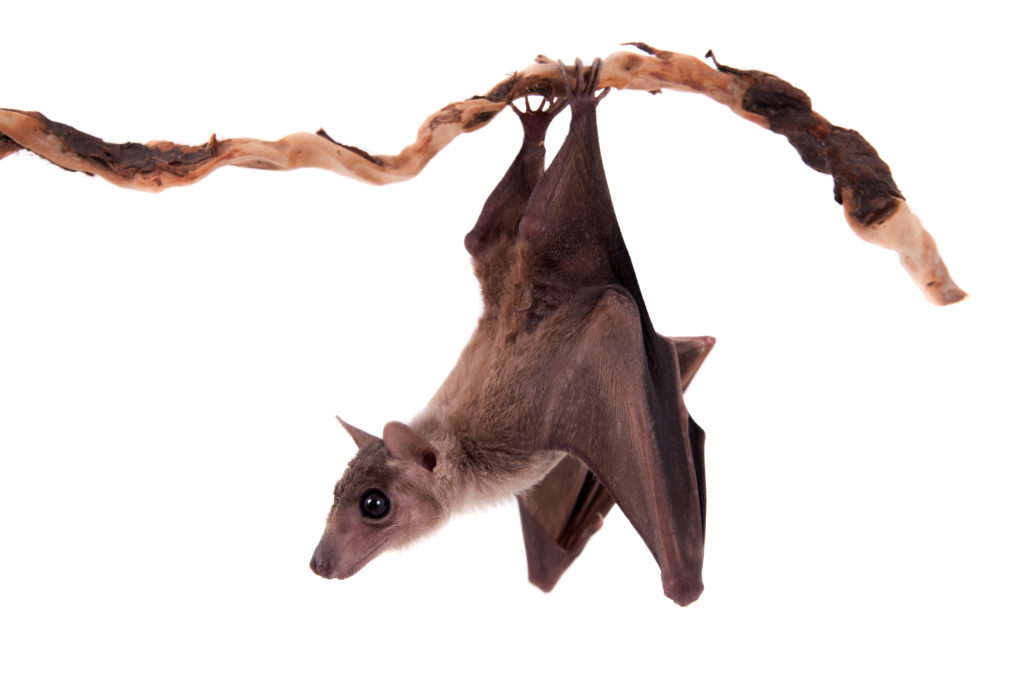
fruit_bat
Měnglà virus was discovered among filoviruses isolated from Old World fruit bats (Rousettus)

All in the viral family

A recent study published in the journal Nature Microbiology provides evidence of a newly identified filovirus species. Using serum samples taken from bats, a well-known host for filoviruses, Yang et al. isolated and identified viral RNA for an unclassified viral genome sequence using next generation sequencing analysis. This new virus genome sequence was organized with the same open reading frames as other filoviruses, encoding for nucleoprotein (NP), viral protein 35 (VP35), VP40, glycoprotein (GP), VP30, VP24, and RNA-dependent RNA polymerase (L). This new genome sequence shared up to 54% of the nucleotide sequences for the filovirus species Lloviu virus (LLOV), EBOV and MARV, with MARV being the most similar. Their analysis suggested that this novel virus should be classified within the Filoviridae family tree as a separate genus, Dianlovirus, and was named Měnglà virus (MLAV).

Continue reading “Meet Měnglà Virus: the newest cousin in the Ebola and Marburg virus family tree”

Over 50 Million Died in the Pandemic of 1918-A Century Later We are still Searching for a Universal Flu Vaccine

One hundred years ago, the world was taking its first deep breaths as it celebrated the end of World War I. The Armistice of Compiègne, was signed on November 11,1918, officially ending the four-year long conflict, which claimed the lives of more than 8 million soldiers (1). What the world didn’t yet realize was that they had been battling a far deadlier enemy in the hospitals and at home than any army the soldiers faced on the fields of war.

During the last year of the war, a deadly influenza virus rampaged around the globe leaving between 50 and 100 million dead in its wake.

Influenza Ward, France 1918. 


The boys were coming in with colds and a headache and they were dead within two or three days. Great big handsome fellows, healthy men, just came in and died. There was no rejoicing in Lille the night of the Armistice.
Sister Catherine Macfie from her post at casualty clearing station no. 11 at St André near Lille, France (2).

Continue reading “Over 50 Million Died in the Pandemic of 1918-A Century Later We are still Searching for a Universal Flu Vaccine”

How To Make Medicine on Mars

Today NASA’s InSight lander will touch down on Mars. InSight, which launched on May 5, is NASA’s first Mars landing since the Curiosity rover in 2012. The lander will begin a two-year mission to study Mars’ deep interior, gathering data that will help scientists understand the formation of rocky planets, including Earth.

NASA's InSight lander approaching Mars.
Image credit: NASA/JPL-Caltech

While every spacecraft that reaches Mars offers more knowledge of the Red Planet, a lot of the excitement is fueled by hopes that someday these missions will bring humans to Mars and enable us to start colonies there. While this goal seems very distant, tremendous progress is being made. Scientists around the globe are making incremental discoveries that will lead to the advances necessary to make colonization of Mars a reality.

I had the pleasure of meeting one team of scientists doing just this—eight high school students from iGEM Team Navarra BG. I met the team and their advisors at the 2018 iGEM Giant Jamboree, where they presented their synthetic biology project, BioGalaxy, as part of the iGEM competition. The problem they aimed to solve is key to helping humans stay on Mars for an extended period of time—how do you take everything you need when there isn’t enough room on the spacecraft? Continue reading “How To Make Medicine on Mars”

A Tale of Two Toxins: the mechanisms of cell death in Clostridium difficile infections

When someone is admitted to a hospital for an illness, the hope is that medical care and treatment will help them them feel better. However, nosocomial infections—infections acquired in a health-care setting—are becoming more prevalent and are associated with an increased mortality rate worldwide. This is largely due to the misuse of antibiotics, allowing some bacteria to become resistant. Furthermore, when an antibiotic wipes out the “good” bacteria that comprise the human microbiome, it leaves a patient vulnerable to opportunistic infections that take advantage of disruptions to the gut microbiota.

One such bacteria, Clostridium difficile, is of growing concern world-wide since it is resistant to many different antibiotics. When a patient is treated with an antibiotic, C. difficile can thrive in the intestinal tract without other bacteria populating the gut. C. difficile infection is the leading cause of antibiotic-associated diarrhea. While symptoms can be mild, aggressive infection can lead to pseudomembranous colitis—a severe inflammation of the colon which can be life-threatening.

C. difficile causes disease by releasing two large toxins, TcdA and TcdB. Understanding the role these toxins play in colonic disease is important for treatment strategies. However, most published research data only report the effects of the toxins independently. A 2016 study demonstrated a method of comparing the toxins side-by-side using the same time points and cell assays to investigate the role each toxin plays in the cell death that leads to disease of the colon. Continue reading “A Tale of Two Toxins: the mechanisms of cell death in Clostridium difficile infections”