Virus-Like Particles: All the Bark, None of the Bite

Globally, there have been over 5 million deaths attributed to COVID-19 since the start of the pandemic. Throughout the ongoing battle against SARS-CoV-2, researchers have been studying the viral lineage and the variants that are emerging as the virus evolves over time. The more opportunities that the virus has to replicate (i.e., the more people it infects), the greater the likelihood that a new variant will emerge.

This short video from the World Health Organization explains how viral variants develop.

The US Centers for Disease Control and Prevention (CDC) classify SARS-CoV-2 variants into four groups: Variants Being Monitored (VBM), Variants of Interest (VOI), Variants of Concern (VOC) and Variants of High Consequence (VOHC). So far, no variants in the US have been identified as VOHC or VOI. Currently, the most common variant in the US is the Delta variant (which includes the B.1.617.2 and AY viral lineages), and it is classified as a VOC.

The Delta variant originated in India and spread rapidly across the UK before making its way into the US (1). Current vaccines, including mRNA and adenoviral vector vaccines, have demonstrated effectiveness against the Delta variant. However, it is a VOC because it is more than twice as contagious as previous variants, and some studies have shown that it is associated with more severe symptoms.

A recent study (2) provides one explanation for the higher infectivity of the Delta variant, using an approach based on virus-like particles (VLPs). The research team was led by Dr. Jennifer Doudna, 2020 Nobel Prize winner for her work on CRISPR-Cas9 gene editing, and Dr. Melanie Ott, director of the Gladstone Institute of Virology at the University of California–Berkeley.

Continue reading “Virus-Like Particles: All the Bark, None of the Bite”

How Can You Improve Protein Digests for Mass Spectrometry Analysis?

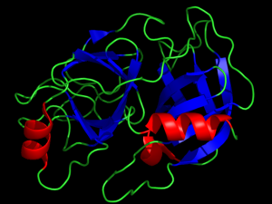
Can predigestion with trypisin (ribbon structure shown) improve protein digests for mass spectrometry analysis?
Can pre-digestion with trypsin improve mass spec analysis?

The trypsin protease cleaves proteins on the carboxyterminus of Arginine (Arg) and Lysine (Lys). This cleavage reaction leaves a positive charge on the C-terminus of the resulting peptide, which enhances mass spectrometry analysis (1,2). Because of this advantage, trypsin has become the most commonly used protease for mass spectrometry analysis. Other proteases which cleave differently from trypsin, yielding complementary data are also used in mass spec analysis: these include Asp-N and Glu-C , which cleave acidic residues, and chymotrypsin which cleaves at aromatic residues. The broad spectrum protease, proteinase K is also used for some proteomic analyses. In a recent study, Dau and colleagues investigated whether sequential digestion with trypsin followed by the complementary proteases could improve protein digests for mass spectrometry analysis.

Continue reading “How Can You Improve Protein Digests for Mass Spectrometry Analysis?”

New Assay to Study SARS-CoV-2 Interaction with Human ACE2 Receptor

Severe acute respiratory syndrome (SARS) is a viral respiratory disease caused by a SARS-associated coronavirus. The most recent version, SARS-CoV-2 was first detected in China in the winter of 2019 and is responsible for the current COVID-19 (coronavirus disease 2019) global pandemic. This virus and its variants have resulted in over 200 million infections and more than 4 million fatalities world-wide. To combat this deadly outbreak the global research community has responded with remarkable swiftness with the development of several vaccines and drug therapies, all produced in record time. In addition to vaccines and drug therapies, diagnostic kits and research reagents continue to roll out to track infections and to help find additional therapies.

This peer-reviewed paper published in Nature Scientific Reports by Alves and colleagues demonstrates how a new assay can be used to discover novel inhibitors that block the binding of SARS-CoV-2 to the human ACE2 receptor as well as study how mutations in the SARS-CoV-2 Spike protein alter its apparent affinity towards human ACE2. The paper also details studies where the assay is used to detect the presence of neutralizing antibodies from both COVID-19 positive samples as well as samples from vaccinated individuals.

Continue reading “New Assay to Study SARS-CoV-2 Interaction with Human ACE2 Receptor”

Illuminating the Kinome: NanoBRET Target Engagement Technology in the Spotlight

Updated November 21, 2023

In the life of a cell, phosphorylation of proteins is an everyday occurrence. The transfer of a phosphate group, from a molecule such as adenosine triphosphate (ATP) to a specific functional group on a protein, is catalyzed by a protein kinase. The vast majority of protein kinases are classified as either serine/threonine kinases or tyrosine kinases; over 500 kinase genes have been identified in the human genome (1).

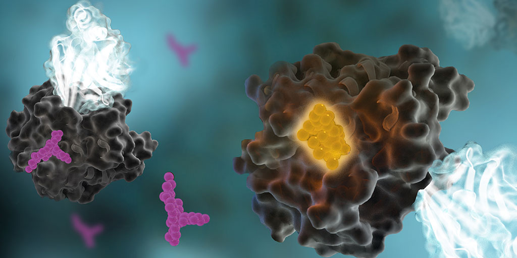
nanobret-Target-Engagement-1024x512-1

Protein phosphorylation is a key step in most cell signaling pathways, in response to external or internal stimuli, and it is not surprising that dysregulation of these pathways contributes to a variety of cancers. The first oncogene to be characterized was SRC, a gene that encodes a tyrosine kinase (reviewed in 2). With more kinases being implicated in oncogenic pathways, significant drug discovery efforts have been devoted to developing and characterizing inhibitors of protein kinases. These efforts have accelerated ever since the first targeted small-molecule kinase inhibitor, imatinib, received US FDA approval in 2001 for the treatment of chronic myeloid leukemia (3). Since that time, many more protein kinase inhibitors have received FDA approval, with 67 small-molecule inhibitors listed as of September 2021.

Continue reading “Illuminating the Kinome: NanoBRET Target Engagement Technology in the Spotlight”

Evidence of Inflammasome Activation in Severe COVID-19

The pandemic caused by SARS-CoV-2 has brought the world to its knees. There have been many deaths, many persons with lingering disease (long COVID) and the inability to vaccinate everyone quickly, for starters. SARS-CoV-2 has not only been a tricky adversary in terms of treatment options to save lives, it’s also been a wily opponent to researchers studying the virus.

Contributing to the existing studies, with their review of the role of inflammasomes in COVID-19, Vora et al. recently published “Inflammasome activation at the crux of severe COVID-19” in Nature Reviews Immunology. In this paper they detail evidence of inflammasome activation and its role in SARS-CoV-2 infections.

Contributions of Those Lost in the SARS-CoV-2 Pandemic
I’d like to take a moment to note the uniquely awful nature of the virus at the center of this blog and the paper it reviews. Many of the papers we blog about describe research involving cell lines, mice or another animal model. The closest most reports get to human research subjects is the use of human cells lines. In the Vora et al. report, serum and tissue samples are from actual human patients, some that survived and many that did not survive COVID-19. It’s not lost on us, Dear Reader, the contributions of those that suffered and died due to SARS-CoV-2 infection. Many persons with severe or fatal COVID-19 have made a significant contribution to our understanding of this virus and its treatment options. We owe them, as well as the researchers that have studied SARS-CoV-2, our sincerest gratitude.

Why the Interest in Inflammasomes?
For detailed information on inflammasomes you can read Ken’s blog, here. You will find background information there and on our inflammasome web page.

In their paper, Vora et al. provide evidence of inflammasome activation, both direct and indirect, in COVID-19. The authors note:

“Key to inflammation and innate immunity, inflammasomes are large, micrometrescale multiprotein cytosolic complexes that assemble in response to pathogen-associated molecular patterns (PAMPs) or damage-associated molecular patterns (DAMPs) and trigger proinflammatory cytokine release as well as pyroptosis, a proinflammatory lytic cell death.”

Continue reading “Evidence of Inflammasome Activation in Severe COVID-19”

New Therapy for Brain Tumors: High-Pressure Oxygen Rewires Glucose Metabolism in Glioblastoma

Glioblastoma (GBM) is an aggressive type of brain tumor, and one of the deadliest cancers. GBM is often treated with surgery, radiation and chemotherapy, but even if the initial treatment is successful, a majority of patients relapse within months. One reason why GBM is so difficult to treat is the hypoxic (low-oxygen) tumor environment. It is known that hypoxic cells are resistant to radiotherapy; the greater the number of tumor stem cells in a hypoxic environment, the less efficient radiotherapy is at controlling tumor growth.

A new therapeutic approach aims to remove the hypoxic environment in GBM by administering pure oxygen to patients at high pressure, known as “hyperbaric oxygen (HBO) therapy”. Previous studies have shown that HBO improves the efficacy of radiotherapy in GBM patients. However, the therapeutic mechanism of HBO was largely unknown. That is, until now.

Dr. Anna Tesei, the Head of Radiobiomics and Drug Discovery at the Biosciences Laboratory of IRST-IRCCS in Italy, recently published a study on the mechanism in which HBOT affects GBM tumor cells and the tumor environment. “The main purpose of our study was to provide a preclinical rationale for the use of hyperbaric oxygen in association with radiotherapy for the treatment of GBM,” she says.

Dr. Anna Tesei is studing glucose metabolism in glioblastoma cells.
Continue reading “New Therapy for Brain Tumors: High-Pressure Oxygen Rewires Glucose Metabolism in Glioblastoma”

LncRNA: The Long and Short of “Junk RNA”

The Central Dogma and Junk DNA

lncRNA, long noncoding RNA

On September 19, 1957, Francis Crick delivered a lecture during a symposium at University College London, titled “Protein Synthesis”. The lecture was published a year later (1); in it, Crick quotes his colleague James Watson as saying, “The most significant thing about the nucleic acids is that we don’t know what they can do.” In contrast, Crick argued that proteins play a central, indispensable role as enzymes within the cell that catalyze a variety of chemical reactions. He believed that the main role of genetic material was to control the synthesis of proteins, although the mechanism of that process was not known.

Crick’s hypothesis came to be known as the central dogma of molecular biology, and it was immortalized in his hand-written notes that described the flow of information from DNA to RNA to proteins. This achievement was all the more remarkable, considering that messenger RNAs were completely unknown at that time, and very little was known about how the cellular translational machinery functioned within the cytoplasm to synthesize proteins. Although the later discovery of retroviruses appeared to challenge Crick’s central dogma, he explained quite succinctly that his original statement had simply been misunderstood, and that information could flow in both directions between DNA and RNA (2).

Continue reading “LncRNA: The Long and Short of “Junk RNA””

The NLRP3 Inflammasome: Flipping the Switch

It’s been just over 10 years since the world lost a pioneering immunologist and biochemist, Dr. Jürg Tschopp. He died tragically during a hiking trip in the Swiss Alps on March 22, 2011. A host of academic journals, including Science, Nature and Cell, paid tribute to Dr. Tschopp with obituaries that highlighted his many accomplishments in the fields of apoptosis and immunology.

In 2002, a team led by Dr. Tschopp at the University of Lausanne, Switzerland, was studying the role of the proinflammatory cytokine interleukin 1 beta (IL-1β). This cytokine is produced in the cytoplasm as an inactive precursor (pro-IL-1β). It is cleaved by caspase-1 to the active form, but the exact process by which caspase-1 itself is activated was unknown at the time. Several members of the caspase family contain a conserved region known as the caspase recruitment domain or CARD, and it was proposed that this domain was essential to caspase activation.

Based on similarity to another protein containing an N-terminal CARD motif (Apaf-1) that is involved in activation of caspase-9, the researchers examined the roles of a family of proteins known as NALP1, NALP2 and NALP3 (1). In particular, they were interested in NALP1, which is involved in the immune response. Unlike Apaf-1, NALP1 contains a CARD motif at the C terminus, while the N terminus contains a related motif known as a pyrin-like domain (PYD). The research team had previously showed that the PYD region of NALP1 interacted with an adapter protein known as PYCARD or ASC, which also contains an N-terminal PYD and C-terminal CARD.

The results of the team’s in vitro binding, activation and immunodetection studies showed that a multi-unit protein complex is responsible for caspase activation, and they called this complex the “inflammasome” (1). It is composed of caspase-1, caspase-5, PYCARD/ASC and NALP1.

Continue reading “The NLRP3 Inflammasome: Flipping the Switch”

RAS-Targeted Drug Discovery: From Challenge to Opportunity

cancer cell, ras-targeted drug discovery

In 1963, Jennifer Harvey was studying Moloney murine leukemia virus (MMLV) at the cancer research department of the London Hospital Research Laboratories. After routine transfers of plasma from MMLV-infected rats to mice, she made an unusual discovery. In addition to the expected leukemia, the mice that received the plasma developed solid tumors (soft-tissue sarcomas), primarily in the spleen (1). A few years later, Werner Kirsten at the University of Chicago observed similar results working with mouse erythroblastosis virus (MEV) (2).

Subsequent research, with the advent of genome sequencing, showed that a cellular rat gene had been incorporated into the viral genome in both cases (3). These genomic sequences contained a mutation later shown to be responsible for the development of sarcomas, and the word “oncogene” became a common part of the vocabulary in cancer publications during the early 1980s (4). Harvey’s discovery led to the naming of the corresponding rat sarcoma oncogene as HRAS, while Kirsten’s related oncogene was named KRAS. Several laboratories, working independently, cloned the human homolog of the viral HRAS gene in 1982 (3). The human KRAS gene was cloned shortly thereafter, as well as a third RAS gene, named NRAS (3). Additional studies showed that a single point mutation in each of these genes led to oncogenic activation, and they have been popular targets for anticancer drug discovery efforts ever since.

Continue reading “RAS-Targeted Drug Discovery: From Challenge to Opportunity”

Antibody Response Differ in Adults, Kids and Potential Cross-Reactive Coronavirus Antibodies

B cell and B cell receptor cartoon. The B cell receptors are important for antibody response.
Drawing of a B cell and the B cell receptor. The receptor shows the characteristic Y shape of an immunoglobulin molecule.

B cells are the immune cells that produce antibodies (immunoglobulins or Ig) to detect intruding pathogens. B cells produce a variety of classes of antibodies. Generally during an immune response to a pathogen, whether viral or bacterial, B cells produce immunoglobulins (Ig) IgM and IgD, and later in the response, IgG and IgA, that are specific to the intruding organism. These Igs capture and aid in neutralizing the pathogen.

Ig classes can be studied by sequencing the B cell receptor (BCR), which binds antigen specifically. BCRs are formed via irreversible gene segment rearrangements of variable, diversity and joining (VDJ) genes. Ig classes can be diversified through somatic hypermutation and class-switch recombination of these gene segments (1).

B cell receptors with high sequence similarity can be found in individuals exposed to the same antigen, demonstrating that antigen exposure can result in similar B cell clones and memory B cells between individuals, both adults and children (1).

However, B cell immune responses can differ between adults and children. For example, children use more B cell clones that form neutralizing antibodies to HIV-1. And children infected with SARS-CoV-2 generally have milder illness than infected adults. SARS-CoV-2-infected children have lower antibody titers to the virus and more IgG-specific response to SARS-CoV-2 spike protein than to the nucleocapsid protein (1). These differences can contribute to faster SARS-CoV-2 clearance and lower viral loads in children versus adults.

Continue reading “Antibody Response Differ in Adults, Kids and Potential Cross-Reactive Coronavirus Antibodies”